
Why Flexible Pelvis Models Make Presentations More Engaging
Increasing the level of student engagement during training strongly correlates to increased effectiveness and efficiency. As a student’s clinical studies advance, so does the level of advancement of their training materials. Realistic and interactive training models can give students a visual and hands-on approach to studying injuries and procedures to create more successful outcomes in future treatment plans. And it isn’t just medical trainees who can benefit from these models. They are also essential to patient education, allowing physical therapists and other specialists to demonstrate different movement patterns.
When it comes to studying specific anatomy like that of the pelvis, using a flexible pelvis model encourages increased retention of surgical lessons and demonstrations while also boosting critical thinking. The Socratic Method of lecturing, which involves dialogue between teachers and students, is effective, but higher student engagement directly stems from more active learning. Using a flexible pelvis model during training will allow students to get a more detailed view and understanding of the pelvic anatomy and how different injuries or ailments can affect it. The same models can also become an important part of working with patients to give them a better understanding of their own anatomy and treatments This can help make future patient examinations and surgical or therapeutic interventions more successful.
What Can a Flexible Pelvis Model Demonstrate?
Flexible pelvis models not only show detailed pelvic anatomy, but they can also show a wider range of kinetic motion of the pelvis and the joints attached to it. This gives residents the most well-rounded knowledge of the pelvis and its functions. The most important aspects of the pelvic anatomy that a flexible model can effectively demonstrate are:
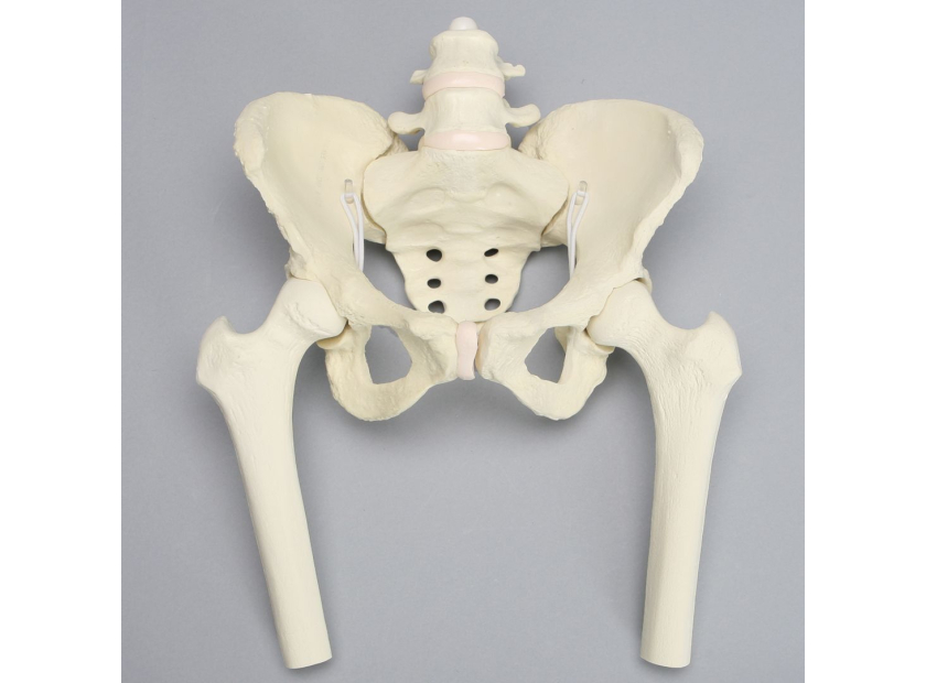
Pelvic Bones
The pelvis is composed of the following three bones that start off as separate bones in childhood and fuse as we reach adulthood:
- Ilium. The ilium is the largest of the three bones. This is the fan-shaped bone on each side of the pelvis laterally.
- Pubis. The pubis is also referred to as the pubic bone. It’s the landmark for pelvic muscle attachment and is located anteriorly and inferiorly—in front of the pelvic girdle.
- Ischium. The ischium is located posteriorly and inferiorly. The inferior part of the ischium, known as the infectious ramus, is the strongest of the hip bones.
In addition to the bones that fuse to create the pelvis, there are two other bones that connect to the pelvis:
- Coccyx. This coccyx is also referred to as the tailbone. It’s a triangular arrangement of bones at the bottom of the spine and is made up of three-five fused vertebrae.
- Sacrum. The sacrum is the triangular-shaped structure located at the base of the lumbar vertebrae to form the posterior pelvic wall. It’s made up of five fused vertebrae.
Pelvic Joints
The pelvis also has several joints that connect the two halves together while connecting the pelvis to the rest of the body:
- Sacroiliac (SI) Joints. The SI joints are made up of the sacrum and connect the two halves of the pelvis on the posterior side of the body. They link the pelvis to the lower spine.
- Acetabulofemoral Joints. These are also known as the hip joints. It’s located between the acetabulum and the femur to connect the pelvis to the lower body and support the weight of the body.
- Pubic Symphysis. The pubic symphysis is centered between the two halves of the pelvis to allow them to articulate with one another. It functions to absorb shock during walking and becomes more flexible to allow a baby to be delivered.
Having a flexible pelvis model can better demonstrate how the two halves of the pelvis move together and independently of one another. This can help students understand the posterior and anterior movements of the acetabulofemoral joints, the forwards and backwards movement from the SI joints, pelvic rotation, and pelvic depression and elevation to easily visualize how the bones and joints work together and make motions such as walking possible.
Types of Flexible Pelvis Models
Flexible Pelvis with Removable Femurs |
Pelvis with Movable SI Joints |
Pelvis with Removable Legs and Movable SI Joints |
Pelvis Attached to Full Spine Model |
|
This model should include flexible SI joints and L4-L5 vertebrae that have semi-flexible discs. Removable femurs bring more buildability to the model for more in-depth demonstrations. |
This model includes the L5 vertebra and the coccyx with movable SI joints. This gives a detailed view of a stand-alone pelvis for more specific demonstrations. |
This model also includes articulating L4-L5 vertebrae with a shock cord to demonstrate posture. Full length femurs, patellae, and proximal tibias with stretch band ligaments give students a more comprehensive view of how the pelvis and surrounding joints work together and their range of motion. |
This model demonstrates C1 to sacrum with a full pelvis and articulates to show spine angular ion and rotation with anterior and posterior ligaments. This can help students understand how spinal deformities affect pelvic rotation and understand treatment options. |
These flexible pelvis models can aid in educating students in specialties such as orthopaedics, chiropractic, physiatry, rheumatology, and pain management.
Demonstrating Pelvis Flexibility for Better Engagement
Increasing the use of flexible pelvis models in demonstrations is the best way to increase knowledge retention in students by allowing them to engage more. The most effective joint models are not only three-dimensional and durable, but they also offer an accurate range of kinetic motion. Being able to see how the pelvis articulates will help impress the knowledge of healthy pelvic movement in students so that they can better diagnose and treat pelvic injuries that could hinder that motion. For patients, the experience of seeing these models demonstrate movement can help them better understand their own conditions and treatments, supporting better outcomes.
The educational models created by Sawbones are designed to make your lessons and your patient interactions more significant and impactful. Contact us online or at 206-463-5551 to learn more.

If you're seeking something you can't find on our website, our sales team is happy to help. We can either direct you to the right model or provide a free quote on the right custom project to meet your needs. Discover options with our clear bone models, laminated blocks, custom displays, or other machining projects.

Work with the industry leader in medical training products!
For four decades, Sawbones has offered the best medical training products for doctors, educators, sales specialists and more. Contact us to learn more about our high-quality models!
1 (206) 463-5551Contact Us!


