
Models for Medical Education
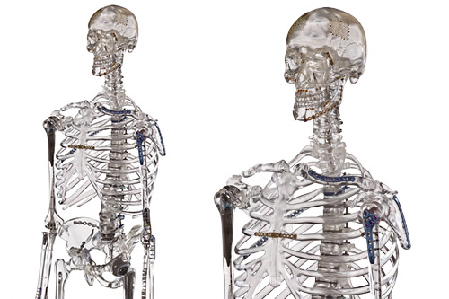
Anatomy models for medical education are taking an increasing role of importance for training institutions today. Cadavers can be hard to source and expensive to manage. The good news is that technology for creating models has advanced to the point where it can replicate many training programs and methods that were once only possible with actual human cadavers.
These models are available for all major segments of the body in varying levels of detail. However, each unique anatomical area has its own specific structure, injury risk, and requirements for treatment. Finding a company capable of providing detailed models for head-to-toe diagnosis and treatment is the most streamlined way to support medical education.
Breaking Down the Human Body into Segments
While the overall human body breaks down into four key components—cells, tissues, organs, and systems—this is a bit simplistic in medicine. Another approach is to separate anatomy into five segments and target treatment based on the unique characteristics of those areas. These five segments are the head, neck, torso, and upper and lower extremities.
Head
|
Neck
|
Torso
|
Upper Extremities
|
Lower Extremities
|
|
The head is comprised of bones and soft tissues that supply blood and nerves while providing protection. |
The neck includes seven cervical vertebrae, portions of the spinal cord, muscles, veins, arteries, and nerves. |
The torso encompasses thoracic and lumbar vertebrae, the abdominal segment, and the pelvic region. |
The upper extremities include the upper arm, forearm, and hand. |
This covers the hip, knee, ankle, and bones of the thigh, leg, and foot. |
Every one of these segments has different levels of complexity, from common injuries physicians encounter often to more complicated issues that require highly specialized treatment. As a result, each has a focus of study instructors should target when looking at the overall structure.
Key Areas of Study: Head
Obviously, the most critical part of the head is the brain—so much so that it's an entire class, and specialty, of its own. Aside from that, several vital bones protect it and support function in the face. Areas of study should target:
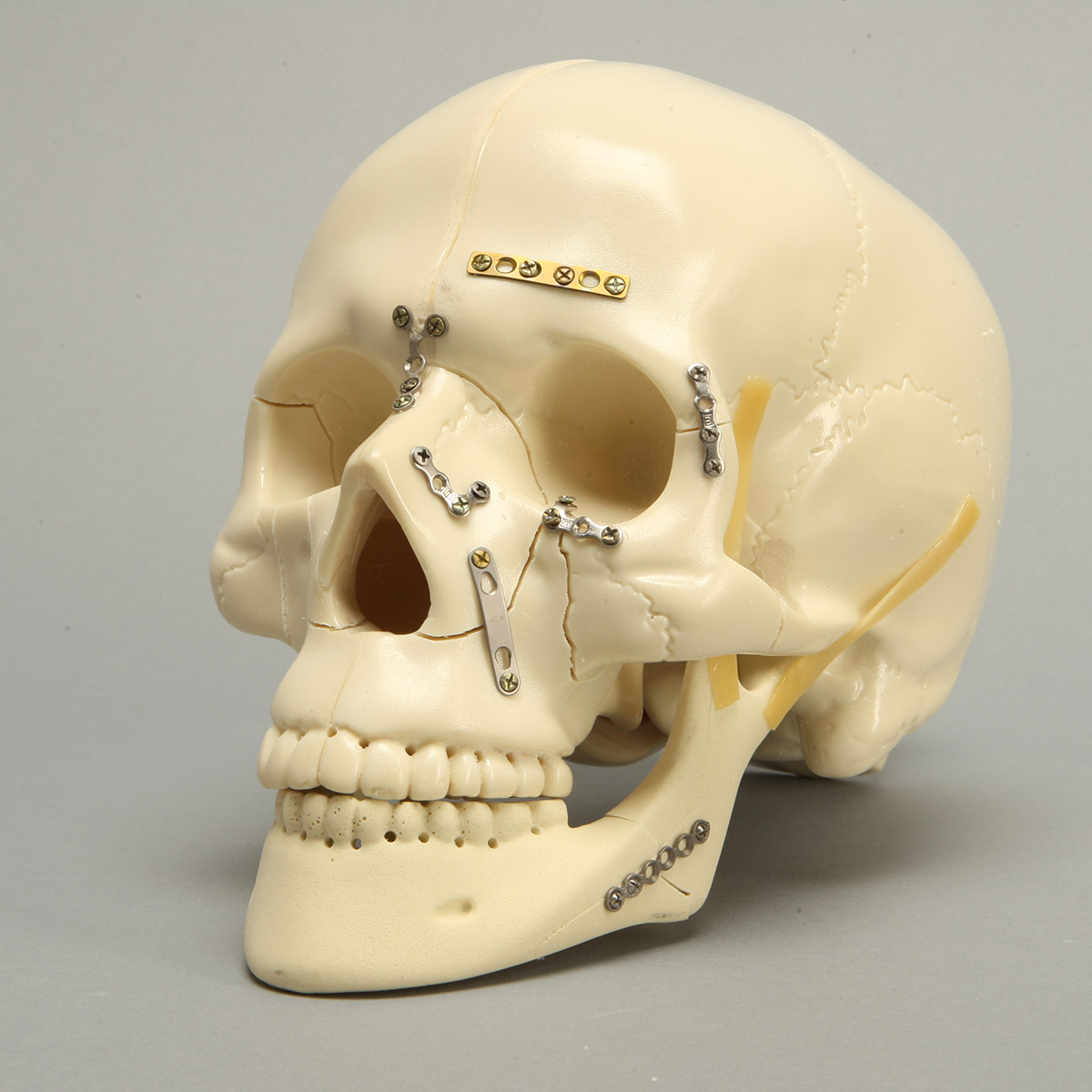
- Cranium: The skull is responsible for the protection of the brain and the head's vascular system. Primary bones include the sphenoid, ethmoid, occipital, frontal, temporal, and parietal.
- Face: The structure supports the face's soft tissue and has 14 bones overall spread out among the maxilla, mandible, zygomatic, nasal, palatine, inferior nasal concha, vomer, and lacrimal areas.
- Auditory: While these bones are located in the cranium, they have their own category because their purpose is to facilitate sounds. This section is composed of three sets of tiny bones: malleus, incus, and stapes.
Damage to any one of these systems is often traumatic and can result in life-threatening circumstances. Doctors may also have to deal with chronic conditions in these areas like tinnitus or temporomandibular joint (TMJ) syndrome.
Key Areas of Study: Neck
The neck has many components designed to both support the head and enable communication and oxygen flow to the body. The list of possible areas of study is virtually endless. Some of the more common focuses include:
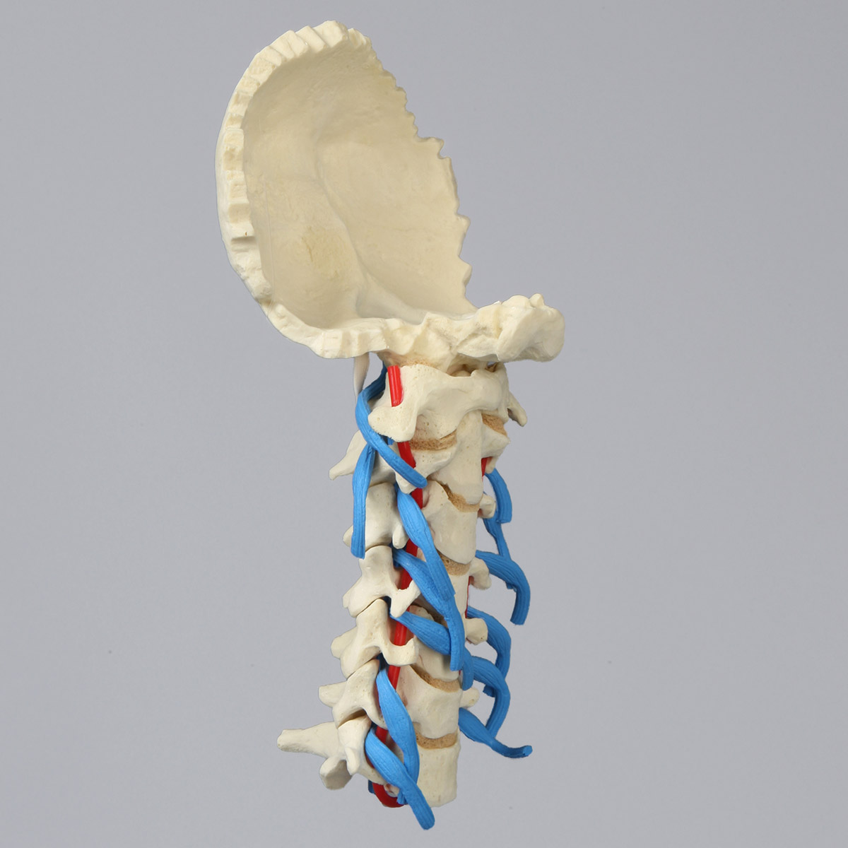
- Cervical vertebrae: There are seven cervical vertebrae in the human body, separated by intervertebral discs made of fibrocartilage. The cervical spine also includes two unique types of joints, the lateral atlantoaxial joints, and the median atlantoaxial joint.
- Jugular veins: These two veins—the internal and external—transport blood from the head back to the heart. They work in conjunction with the superior vena cava in the delivery of oxygenated blood.
- Carotid arteries: Carotid arteries do the opposite of what the jugular veins do. They deliver blood from the heart to the brain and head.
- Larynx: The larynx is a passage that provides air to the lungs. It also holds the vocal cords.
- Vocal cords: These are small, smooth muscles that vibrate and make it possible to speak.
Injuries in the neck are varied. Plaque can build up in the carotid arteries, resulting in carotid artery disease that leads to stroke. Damage to the vocal folds can create vocal cord disorder that impacts the ability to speak. Injuries to the cervical vertebrae create chronic pain and dysfunction. Just about any traumatic injury in this area can be life-threatening.
Key Areas of Study: Torso
The torso is a huge segment of the body and houses the majority of organs, so it will require extensive medical training. This is especially true when considering studies of the spine. Significant areas of focus will include:
- Major organs and systems: The majority of major organs and systems are housed in the torso, including the heart, lungs, kidneys, liver, stomach, and reproductive organs.
- Muscle groups: The torso is home to the pectoral, abdominal, lateral, and epaxial muscles.
- Lumbar spine: There are five lumbar vertebrae, which are the largest in the spine due to their location and stress level. They are joined by intervertebral discs, nerves, muscles, and ligaments.
- Thoracic spine: There are twelve thoracic vertebrae located in the center of the spine. It's also the attachment point for the ribs which protect the lungs and heart.
- Nerves: The spinal cord runs through the torso and is responsible for providing sensation to the skin and sending signals throughout the body.
As the torso houses so many components, any injury to this area is hazardous. It's also home to many chronic pain conditions, especially in consideration of the lumbar spine, where herniations of discs are common.
Key Areas of Study: Upper Extremities
Arms and hands can be surprisingly complex due to the high number of small, interlinking connections in this area of the body. The human hand alone includes many different bones and joints and more than 100 ligaments and tendons. Specific lesson plans may cover:
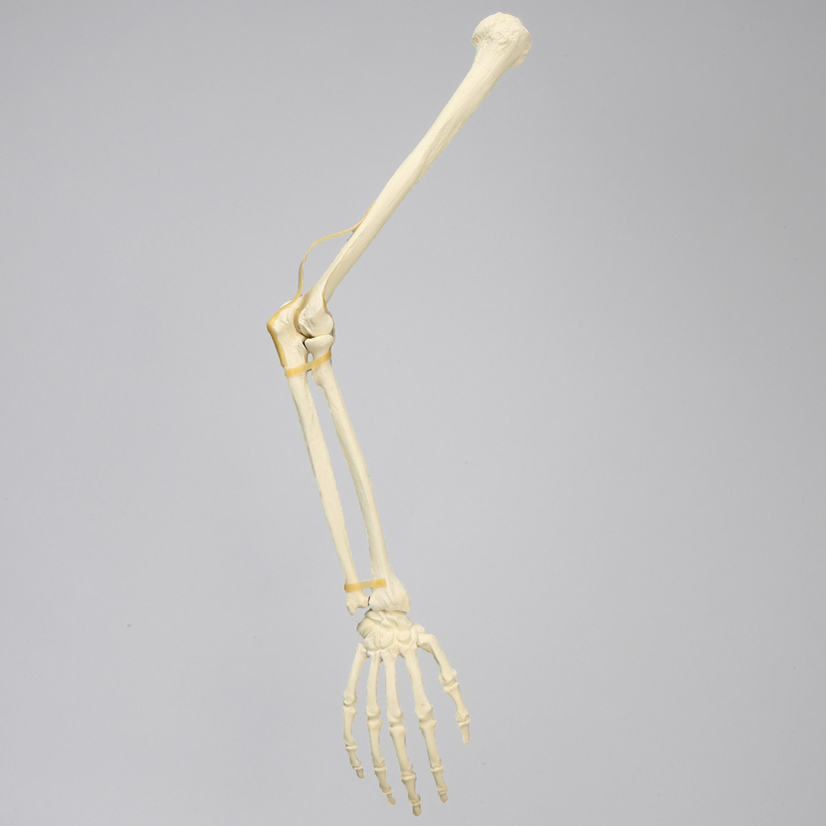
- Shoulder and arm: These areas are comprised of 10 bones grouped by the clavicle, scapula, humerus, ulna, and radius.
- Wrist: The wrist has 16 bones grouped by the pisiform, trapezium, trapezoid, capitate, hamate, scaphoid, lunate, and triquetrum.
- Hand: The hand has the most separate bones of any other part in the upper extremities, including 38 bones broken down to metacarpal and phalanges.
- Joints: The arm has some unique joints, including the ball and socket shoulder joint and the elbow, often referred to as a hinge joint.
Of course, the arm has many different ligaments, vessels, and nerve endings necessary for function. Injuries are common in this region due to regular movement and stress.
Key Areas of Study: Lower Extremities
The lower extremities are under a lot of pressure because they support the weight of the upper body. Injuries in these areas are expected, either as a result of trauma or just aging. Future doctors may find themselves treating conditions related to several areas prone to injury:
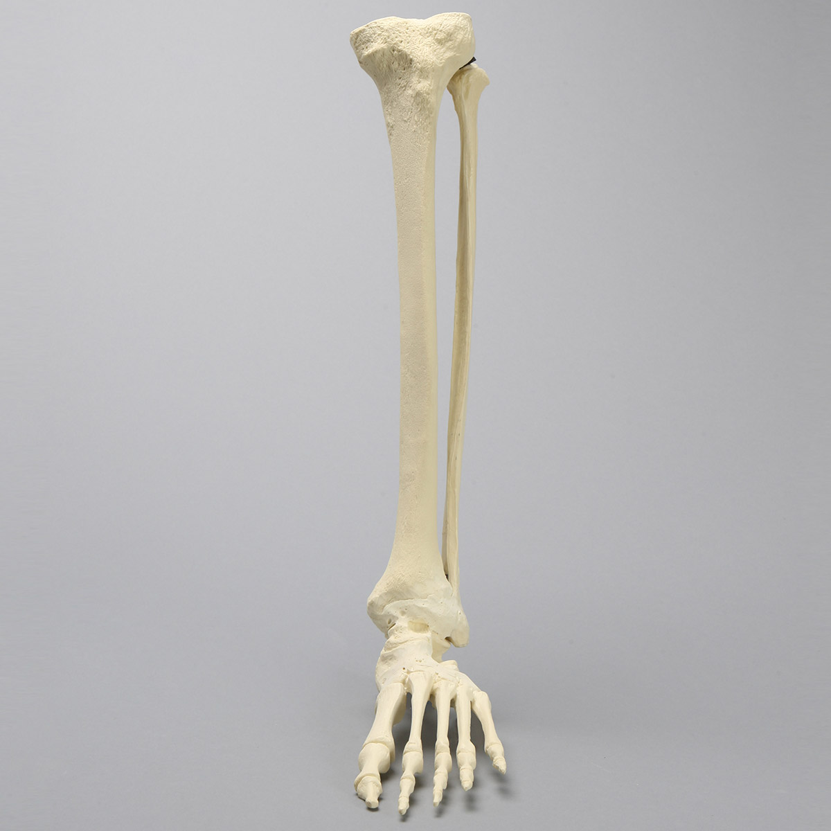
- Innominate bones: The innominate bone refers to the hip because, originally, this very important segment was not given a name. Innominate means "not named or classified."
- Femur: The femur is the longest bone in the human body, making up the thigh structure. It's broken down to the proximal, shaft, and distal components.
- Patella: The patella, or kneecap, connects to the femur and protects the knee joint. It's the largest sesamoid bone—or bone embedded with muscle or tendon—in the body.
- Tibia and fibula: These two bones provide structure to the lower leg, with the tibia in front as the shin and the fibula behind as the calf.
- Tarsals: These bones make up the mid and rearfoot and include the talus, cuniforms, navicular, cuboid, and calcaneus.
- Metatarsals and phalanges: These bones make up the front of the foot, including the toes.
- Muscles: There are muscle groups for the hip, upper leg, lower leg, and foot, all of which provide function and flexion.
- Ligaments and tendons: All of these important components are connected by a series of ligaments and tendons that maintain the structure and provide for movement.
- Femoral artery: This artery, located in the front of the thigh, provides the main blood supply to the leg.
- Sciatic nerve: This nerve runs from the lower back down each leg.
Complications in the lower body often occur due to direct injury or issues with the torso. A good example of this is sciatica, where a herniated disc in the lumbar spine impinges on a nerve and creates radiating numbness and pain. As the cause of the pain may not be directly related to an injury to the lower extremities, diagnosis can be challenging.
Selecting Anatomy Models for Medical Education by Segment
Anatomy models for medical education must be highly detailed to provide residents with the information they need to offer treatment in the future. When breaking it down into various bodily segments, it is wise to look for specific attributes and optional features.
What to look for |
Optional Features |
Sawbones products |
|
Head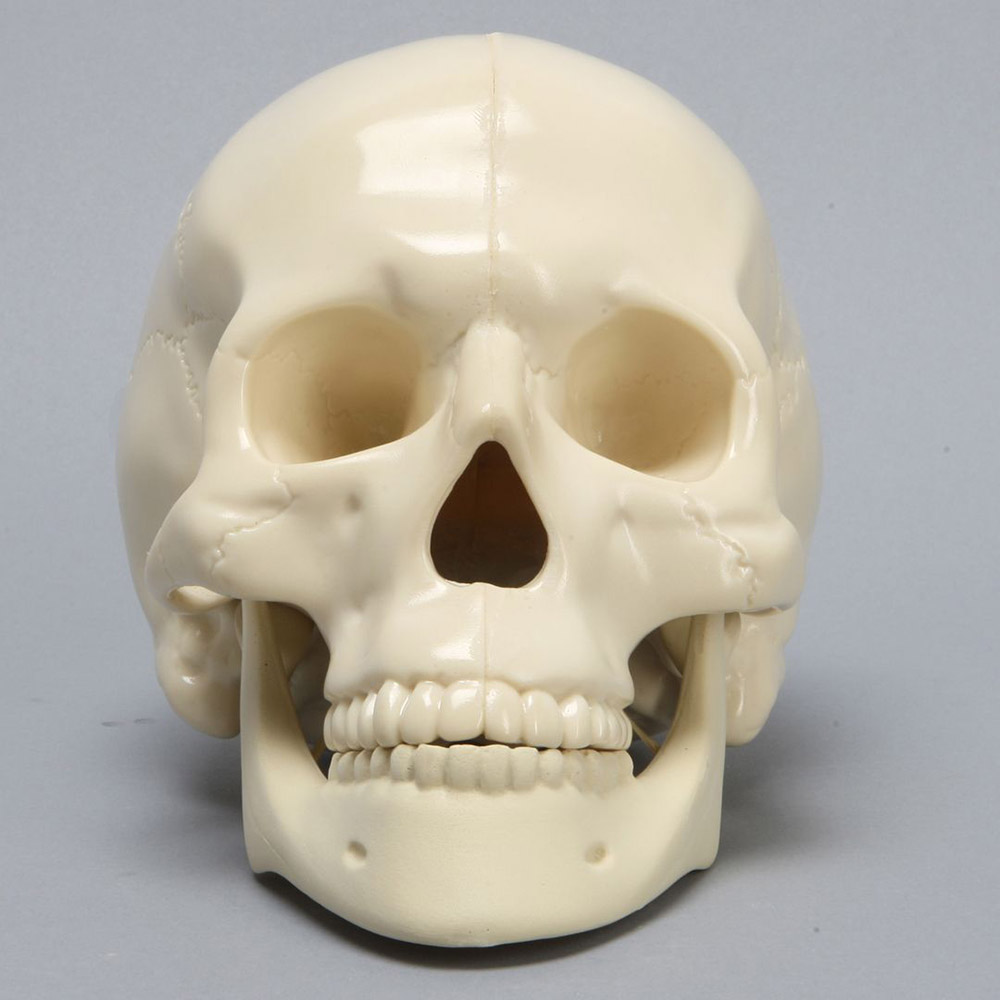 |
The ability to separate the skull into various components is ideal, as it provides a more in-depth study. Vise attachments can also help to affix the model to a workspace. | Removable, replaceable skin is an excellent feature to have, as it allows the student to connect what they've learned about the structure of the face to how it applies to soft tissue. | CMF Specialty Models |
Neck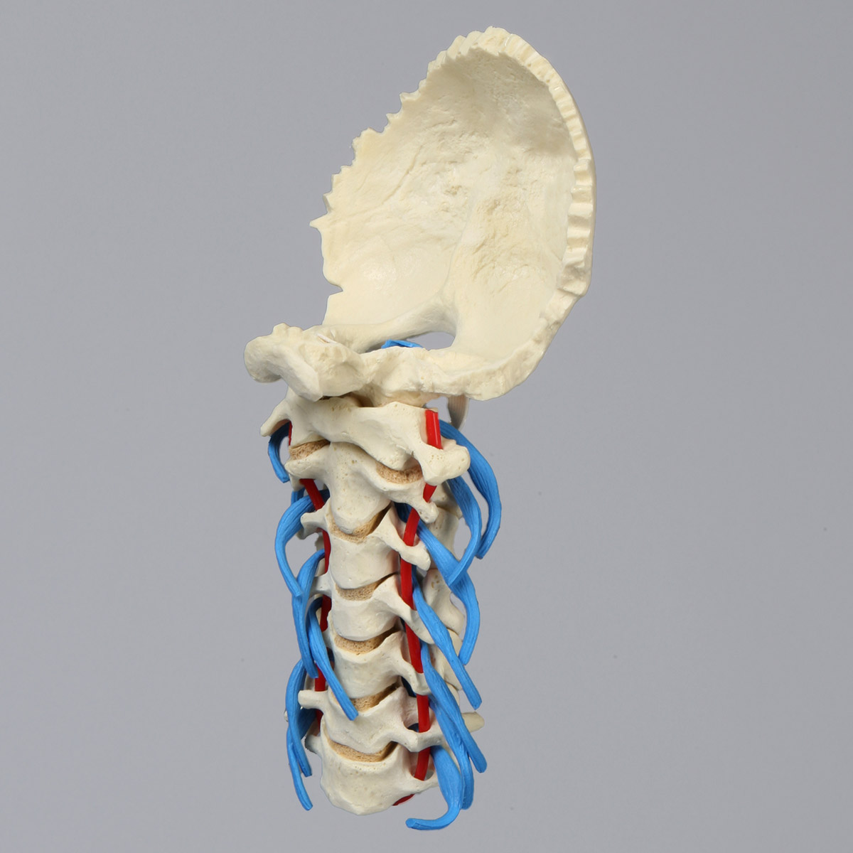 |
Detail is important in the cervical spine, especially regarding function. The vertebrae should move similarly to human motion to help trainees understand kinetic effect. | Contrast can be helpful in guiding trainees on how the discs, ligaments, and nerves intersect the cervical spine and impact other parts of the body. | Sawbones Cervical Models |
Torso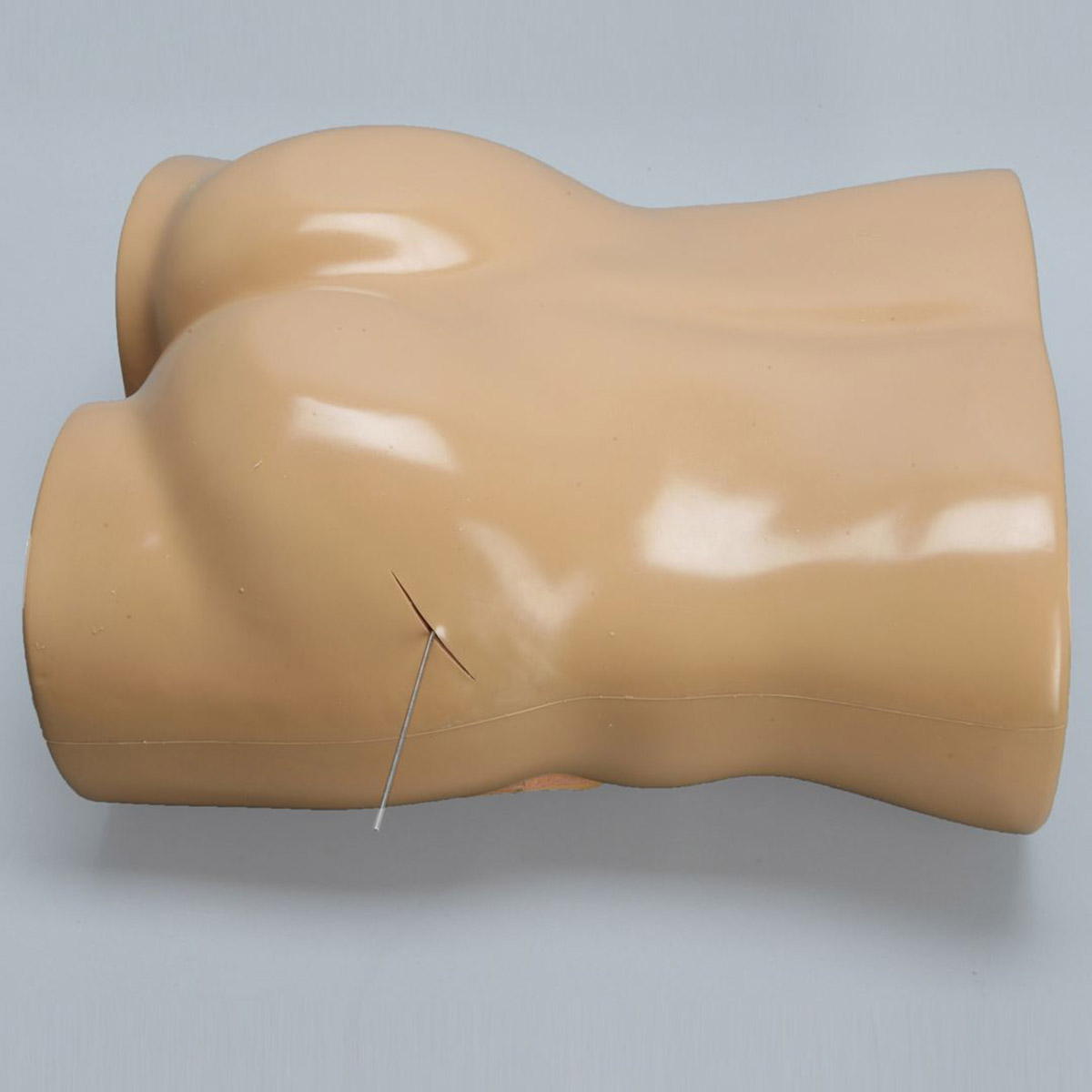 |
Torso models with full spines are ideal, as they allow the student to connect the thoracic and lumbar vertebrae to their overall location. Haptic feedback is also good for injection training. | Radiopaque properties are especially suitable for torso models because they help in medical imaging. Radiopaque material will show up on an x-ray, facilitating the diagnosis of conditions. | Sawbones Torso Models |
Upper Extremities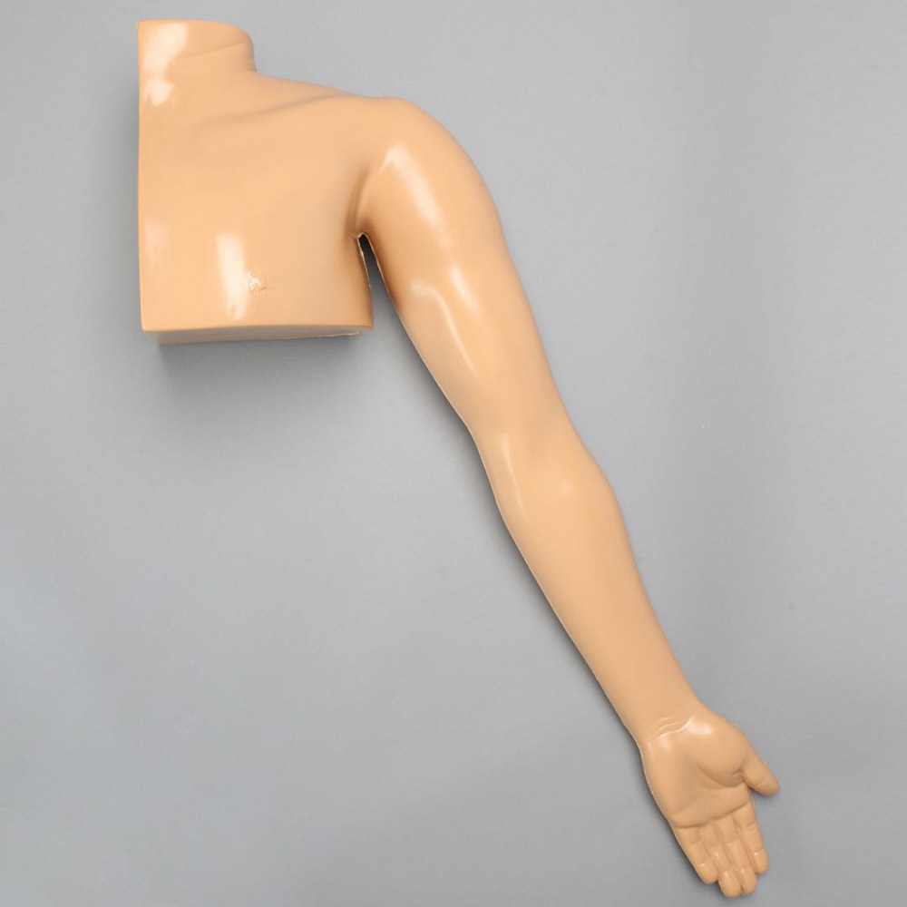 |
Upper extremity models are another area where haptic feedback is ideal for training on injections and other treatments. | Customizable bone fracture options help medical educators show their students various injuries and how they appear in the bones, sharpening diagnostic skills. | Sawbones Arm Models |
Lower Extremities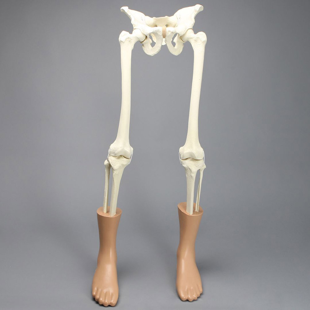 |
Removable pieces show the specific components and how they fit together. Contrasting nerves, tendons, and muscles help trainees better understand the connections. | Special features like haptic feedback, removable skin, and customized fractures are ideal for learning. Arthroscopic training models can also be advantageous here, as this area is a common target for keyhole surgeries. | Sawbones Leg Models |
The best anatomy models for medical education are going to break down components to a granular level. With the right detail, they can replace the need for training on cadavers which can be expensive and impractical. Good medical models provide a head-to-toe learning experience for trainees seeking a well-rounded education.
Sawbones offers a wide range of anatomy models for medical training that support all parts of the body. Visit our contact page or call us at (206) 463-5551 for more information.
