
Effective Knee Synovial Biopsy Techniques Can Be More Successfully Trained With a Bone Model
Minimally invasive procedures are gradually becoming a more preferred standard of treatment, since they are known to significantly reduce the risk of complications and post-operative recovery time. Research has shown that procedures using small incisions for arthroscopy or needle injections for arthrocentesis are taught mostly by having students read published material or wait for a cadaver to be available to practice on. While these have done a decent job of supplementing operating room training, using training models has been proven to bring more successful patient outcomes.
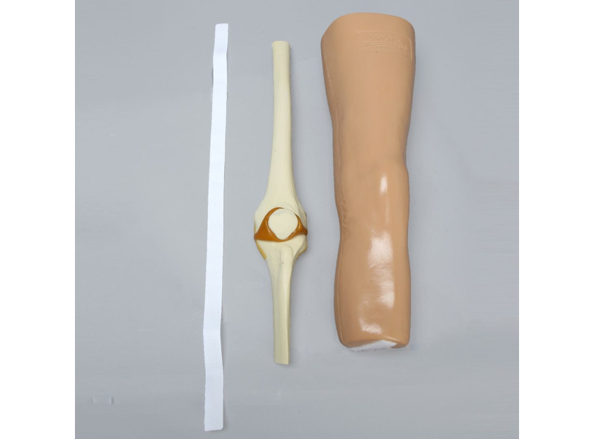
For example, a synovial biopsy of the knee is a minimally invasive procedure performed by rheumatologists or orthopaedic surgeons to remove a sample tissue from the synovial membrane for examination. This is done to help diagnose arthritis, autoimmune disorders, infections, cancer, or other inflammatory conditions. Techniques for procedures like knee synovial biopsy can utilize knee bone models to give residents improved muscle memory for the procedure and reduced surgical error.
Types of Knee Synovial Biopsy Techniques
There are two main ways that a synovial biopsy of the knee is performed:
Arthrocentesis |
Arthroscopy |
|
This is also known as joint aspiration. It is performed by inserting a needle into the joint to abstract synovial fluid from the inflamed area to be sent for testing to determine the diagnosis. An ultrasound may be required to help navigate the needle. |
This is done by making a small incision near the joint and inserting an arthroscope to view the joint while inserting other devices, usually a trocar and biopsy grasper, to cut a small piece of the tissue to be sent for testing. |
Ultrasound-guided synovial biopsies are now performed routinely, with failed biopsies usually being reported as caused by surgical inexperience. Using bone models to simulate a synovial biopsy can help eliminate this by giving trainees an accessible and realistic approach to practice the techniques.
Bone Model Training Improves Technique
Using bone models to train synovial biopsy techniques allows for more exposure and repetition of the procedure, which is known to bring more confidence to those performing it. Having a realistic model that’s durable enough for repeatability is the most effective way to build surgical competency. Bone models for arthrocentesis and arthroscopy help improve technique by:
- Increasing muscle memory to guide the needle and pull fluid from the correct location
- Improving camera and instrument navigation through the joint
- Reducing procedure time
- Reducing the possibility of nerve damage from instruments
- Helping trainees discern what real fluid looks like to ensure proper removal
- Increasing surgical precision when cutting tissue
These procedural benefits have an even larger return on investment when compared with the cost-effectiveness of using a durable bone model that can withstand multiple training sessions and students. Using a bone model that has the advanced features known to provide more efficiency and engagement in lessons can provide residents with their best chance at optimizing future procedure outcomes.
Four Features to Consider for Bone Models
Consider these features when choosing a bone model to help students learn knee synovial biopsy techniques:
- Cosmetic Realism: Bone models with soft tissue and skin can offer a better understanding of where to make the incisions and insert the instruments in a real knee joint. It reduces their chances of damaging nerves or surrounding tissue while maneuvering through the joint and increases their likelihood of success.
- Haptic Feedback: Haptic feedback can gives a better idea of what to expect during arthrocentesis or arthroscopy. Real-time feedback can help steer trainees to the precise location each time and improve the precision of their movements and decision-making in future procedures.
- Kinetic Realism: A realistic range of motion can show how inflammation affects joint movement when deciding on a knee synovial biopsy as a treatment plan. It also allows trainees to experience performing the procedure in a more realistic environment where the joint moves instead of being completely rigid.
- Durability: The most cost-effective way to give students more access to learning the procedure with a bone model, is to consider a model built from durable, reusable materials. This extends the life of the model and allows trainees to practice more to increase their confidence when performing the procedure on patients.
Using a bone model with the proper features for either technique can provide a more practical and economical training route than virtual reality or cadavers. Students can practice as often as they need to in a safe setting before moving on to an operating room setting. Learning knee synovial biopsy techniques from a bone model allows trainees to make mistakes on a realistic joint model and understand how to avoid them in patients. In-depth training with detailed bone models can mean more precise training and positive surgical outcomes.
Sawbones offers advanced knee models to help simulate arthrocentesis and arthroscopic procedures. For more information on our products or to discuss custom training models, contact us at 206-463-5551.

If you're seeking something you can't find on our website, our sales team is happy to help. We can either direct you to the right model or provide a free quote on the right custom project to meet your needs. Discover options with our clear bone models, laminated blocks, custom displays, or other machining projects.

Work with the industry leader in medical training products!
For four decades, Sawbones has offered the best medical training products for doctors, educators, sales specialists and more. Contact us to learn more about our high-quality models!
1 (206) 463-5551Contact Us!


