
5 Reasons a Pelvis Model with Muscles Is Your Best Choice
No part of the human body exists in isolation. Every bone, muscle, organ, and joint operates in concert with the rest of the body, either in immediate connection or through complex networks and balances. This concept is central to a medical student’s core understanding of their future craft and the primary reason why a pelvis model with muscles is the best choice when sourcing training materials.
While there are reasons for using a limited anatomical pelvic unit—such as recreating specific deformities or presentations—choosing a model with muscles and other refinements gives greater context to the structure’s role, movement, and connection to the rest of the body.
A pelvis model with muscles provides:
- Context for the cause of pelvic deformity
- Visual aid for understanding backache
- Clear description of the relationship between the pelvis and spine
- Heightened anatomical understanding
- More effective transfer of core clinical skills
As the pelvis is the fulcrum through which the body’s weight is transferred to the lower limbs in locomotion, a working understanding of the structure has to include the dynamic connection of muscles and joints. When it comes to the central hubs of human anatomy, detail and functionality are essential to successful interventions.
Materials That Promote Understanding
Of course, in order to be of use to modern instructors and their trainees, the muscle-bound pelvic model must authentically recreate the look, feel, and function of the muscles that attach to the structure.
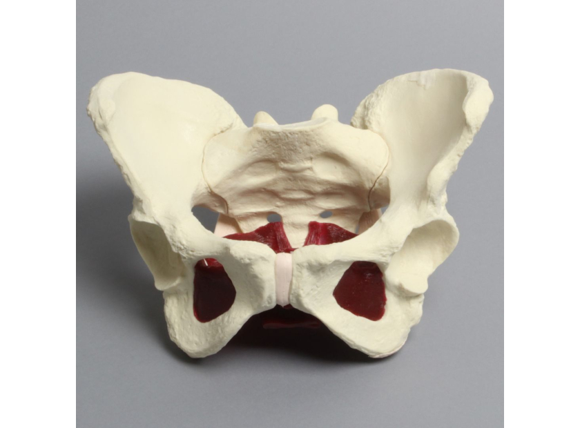
The latest generation of engineered medical training models are designed with just such cutting-edge features. The best pelvis models with muscles use solid foam materials with realistic surface definition and attachment points to lay bare the powerful forces that control movement. These models also contain proportional and realistic vertebrae, discs, moveable muscles, and joints that reveal how the major muscles like the psoas interact with the skeleton at dozens of points both above and below the pelvic girdle.
These materials can be combined with a range of dynamic features to make instruction even more accurate.
Dynamic Features for Every Discipline
Such is the central location of the pelvis that professionals from a range of different medical fields benefit from a working knowledge of its anatomy and performance. By altering the dimensions and scope of an instructional model, teachers can demonstrate conditions and procedures associated with pediatrics, orthopaedics, pain management, neurosurgery, chiropractic, and more.
Such models can be further enhanced with advanced features that provide extra stimulus to non-clinical settings. These features include:
- Buildability for adaptable, multi-purpose models
- Haptic feedback that provides an audio response on contact
- Radiopaque materials for medical imagery training
- Dual open and closed models
- Transparent models for three-dimensional exploration
These features take a model beyond mere anatomical value and provide a more vibrant setting that better prepares students for clinical performance. Their presence is the reason why a pelvis model with muscles, for example, can be more impactful than a static skeletal depiction.
1. Context for the Cause of Pelvic Deformity
An authentic recreation of the sheer size of the muscles connected to the pelvic area provides an easy-to-comprehend visualization of the functional dynamics of the region. The more detailed the model, the more opportunity there is for instructors to convey the range of causes that contribute to pelvic deformity, pain, and abnormality.
2. Visual Aid for Understanding Backache
Adding features such as the psoas muscle to a pelvic model allows instructors to isolate the practical causes of structural abnormalities and deformity. The powerful muscles that run across the spine and pelvis are often the cause of back pain—which is among the most common presentations, impacting an estimated 65 million Americans. Realistic points of attachment and authentic recreations of the scale of muscle-to-bone make clear the forces at play through the region.
3. Clear Description of the Relationship Between the Pelvis and Spine
The movement and strength of the pelvic region rests not on the skeletal connections with the spine but the broad muscles that envelop both structures. Only a model with detailed and realistic muscle groups can convey the interplay between the two most important sites of human movement.
Similarly, there are full spinal models available that authentically recreate the range of movement within the column, and the complications that arise when this motion is restricted.
4. Heightened Anatomical Understanding
All motion through the pelvis is dependent on the joints and muscles of the region. These components pivot and leverage from adjoining skeletal features to provide force. A model that lacks the definition of such muscles does not fully convey the complicated anatomical structure of the area or prepare trainees for their navigation during invasive procedures, especially those involving screws and plates.
Advanced pelvic models can include a complete network of the gluteus maximus, medius, and minimus, rectus femoris, obturator internus and externus, piriformis, sartorius, quadratus femoris, and facia lata muscles around a hip joint.
5. More Effective Transfer of Core Clinical Skills
The goal of heightened realism and detail in a pelvis model with muscles is to prepare trainees for clinical settings. Studies have found that advanced benchtop and engineered models are effective at transferring core skills to the operating room. This transformation can result in shorter procedures, fewer errors, and better surgical communication and leadership.
The continued advancement in the quality and dynamic functionality of medical training models gets students closer and closer to clinical settings while removing the cost and management of cadaveric solutions, and the dangers and limited presentations of operating room experience.
Importantly, the designers of these medical training models are now available to partner with leading educators toward the creation of still more effective teaching tools.
A Pelvis Model with Muscles Is Just the Beginning
Today’s leading medical training model engineers can apply their talents to the creation of training materials for any speciality, and in reproduction of any presentation or procedure.
There are already a number of successful custom models currently being commercially produced to aid doctors and educators around the world. These models are the direct result of practical collaborations designed to improve educational settings.
If adding muscles to a pelvic model seems like just the start of what’s required to better instruct medical professionals, then it’s time to consult a design expert.
The engineers at Sawbones can produce a medical training model of the scale, functionality, and complexity that improves the way you teach your course. For more information on our offerings or to talk about custom training models, contact us at 206-463-5551.

If you're seeking something you can't find on our website, our sales team is happy to help. We can either direct you to the right model or provide a free quote on the right custom project to meet your needs. Discover options with our clear bone models, laminated blocks, custom displays, or other machining projects.

Work with the industry leader in medical training products!
For four decades, Sawbones has offered the best medical training products for doctors, educators, sales specialists and more. Contact us to learn more about our high-quality models!
1 (206) 463-5551Contact Us!


