
Using a Femur Deformity Model to Demonstrate FAI and Other Abnormalities
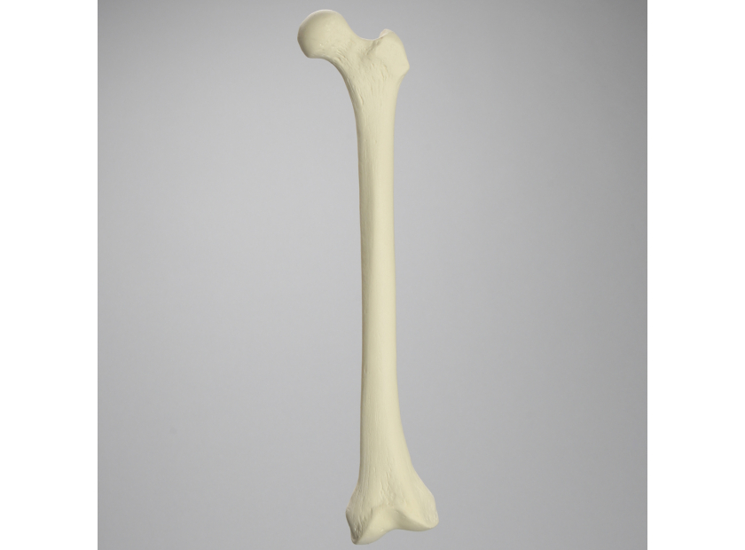
The subtleties and complexities of femur deformity, from femoroacetabular impingement to the full range of shaft fractures, can make it seem that instruction and patient education must always be a matter of approximation. But modern medical models are catching up to the needs of the industry.
Advances in materials, design accuracy, and functionality have led to the creation of a range of femur deformity models that authentically replicate femoral and acetabular lesions, varying degrees of distal valgus deformity and rotation, neck deformities, cortex wall loss, and other common presentations. These models can effectively be used to demonstrate best practices to trainees or educate patients about their conditions.
A Common Enemy
The need for accurate femur deformity models stems from the commonality of conditions such as femoroacetabular impingement. A recent study of more than 1100 hips found mixed femoral and acetabular lesions in around 45% of cases. These painful growths are especially concerning for their close association with hip disorders and the onset of arthritis.
Treatments vary across age groups, the specifics of presentation, and the expectations of the patient—athletes have needs unique to their discipline, for example, while there is continuing debate about the benefits of open versus arthroscopic interventions for femoroacetabular impingement. Each presiding specialist, however, has to begin their exploration of this notoriously difficult-to-diagnose series of disorders in the abstract.
To benefit from the hands-on nature of today’s medical education, they need access to femur deformity models that accurately recreate real-world conditions.
Why Models Work Better
Until the 1990s, surgical procedures were taught through observation. In the 1950s, the only available resource was the operating room. Beginning in the 1960s and improving in quality throughout the 1980s, students were given access to instructional videos.
Today, medical instructors have a range of simulation models at their disposal to help reduce the steep learning curve associated with modern surgical equipment, such as arthroscopes and robots.
Engineered models with advanced materials offer several key advantages to today’s medical training institutions. A femur deformity model, for example, has the following strengths:
- A perfect view – Abstract models isolate the deformity from surrounding tissues to give the student, or patient, a clear understanding of the presentation.
- Robust, reusable materials – Unlike cadaveric models or clinical presentations, models can withstand repeated use and can be altered in line with developing skills.
- Purpose-built – A customized design gives the instructor complete control over the specific presentation and the desired student interaction.
- Realistic materials – Unlike virtual simulations, an engineered solution is built of lifelike composite materials that recreate the feel and performance of real human bone.
- Enhanced diagnostic skills training – Students exposed to a set of femur deformity models better understand how to diagnose injuries and growths and the appropriate intervention for each presentation.
The evolution of medical training models over recent years means that femur deformity models are available and accurate, ultimately improving patient outcomes.
Deformities by Design
It is possible to create medical training models specific to any newly established or previously under-resourced procedure or condition. Such a process is already underway with femur deformity models, and has resulted in an array of different training and patient options.
Femur with a 10, 20, 25, or 30 degree distal valgus deformity
Valgus knee deformity is typically defined as having a femoral angle greater than 10 degrees. This series of models demonstrates the deformity through advancing degrees of severity. The models are made from a rigid foam shell with inner cancellous material that makes cutting and drilling easier. There are also models available that include the knee joint and tibia.
Femur with cortex wall loss
This model details the challenges of hip reconstruction with regard to the integrity of the femur walls. In this instance, a Paprosky Type 2C deformity exists, which means moderate bone loss and deficient walls but with intact acetabular columns. The superior dome is also intact, preventing vertical displacement, but such presentations have a deficient medial wall causing direct medial migration of the hip center. The model includes lifelike cancellous inner materials to better prepare students for the operating room.
Radiopaque femur
Radiopaque materials absorb x-rays meaning these femur models can be used to recreate femur deformity imaging and diagnosis. The varying degrees and subtle variation of femoral and acetabular lesions in femoroacetabular impingement make any recreation of the diagnostic process valuable.
Transparent femur models
Highlighting the potential of abstract resources, these transparent models give students a literal insight into femur construction. Available within a range of constructions, these models feature authentic cancellous inner materials that replicate the porous network within bones with a visual element.
These transparent models demonstrate how engineered solutions can move beyond the constraints of cadavers and the operating room to present instructors and medical professionals with practical resources that improve the efficiency of their demonstrations and courses.
Rather than tethering students to only observing live operations, as was the case 60 years ago, today’s resources bring authenticity, impact, and transferable skills to classrooms where hands-on repetition can safely and affordably teach core skills.
Create the Femur Deformity Model You Need
The medical training models described above are the result of collaboration between expert engineers and working professionals. They are a combination of technology and expertise that is designed to have practical application in teaching the next generation of specialists.
The subtleties of femur deformity and the complex interventions and therapies that follow from their diagnosis can be effectively captured in teaching models, and solutions are available or can be engineered to suit any field or instructional need.
Sawbones can create authentic teaching and demonstration models to support any course or specialty, across any field. For more information on our offerings or to talk about custom training models, contact us at 206-463-5551.
If you're seeking something you can't find on our website, our sales team is happy to help. We can either direct you to the right model or provide a free quote on the right custom project to meet your needs. Discover options with our clear bone models, laminated blocks, custom displays, or other machining projects.

Work with the industry leader in medical training products!
For four decades, Sawbones has offered the best medical training products for doctors, educators, sales specialists and more. Contact us to learn more about our high-quality models!
1 (206) 463-5551Contact Us!


