
Why a Shoulder Joint Model with Ligaments Is the Best Choice for Labrum Repair Training
The labrum is cartilage that surrounds the shoulder joint to act as the attachment site and allows the upper arm bone—the humerus—to fit in the shoulder socket and move freely within it. Through overuse, injuries, and aging processes, the labrum can become brittle and tear, causing reduced shoulder movement and intense pain. Labral repair surgery is the procedure performed to treat the labrum tear and regain easy motion of the shoulder joint without pain.
Common Labral Tears and Repairs
The two most prevalent labral tears are SLAP (superior labrum from anterior to posterior) tears and Bankart tears.
SLAP Tear
A SLAP tear occurs at the front of the upper arm where the bicep muscle tendon connects to the labrum and runs to the back in the shoulder socket. It’s most common in athletes with chronic overhead or snap movements. It results in reduced arm movement as well as pain centralized more at the front of the socket near the bicep tendon.
The SLAP repair is done arthroscopically, which is a minimally invasive technique using an arthroscope inserted into the shoulder through small incisions to inspect the labral tear. If the tear solely affects the labrum, then the torn area of the labrum will be removed. If the tear includes the bicep tendon, anchored sutures will be used to reattach the torn labrum tendon to the bone.
Bankart Tear
A Bankart tear is also known as a Bankart lesion. It commonly occurs when a person dislocates their shoulder. When the shoulder joint separates from the socket, the tissues that surround it can pull the lower front quadrant of the labrum—the glenoid labrum—and tear it. This greatly reduces the shoulder joint’s stability and shock absorption, leading to a higher recurrence of shoulder dislocations.
A Bankart repair is done by reattaching the joint capsule to the glenoid labrum with sutures. This is done to reintroduce stability to the anterior shoulder and help prevent future dislocations.
Posterior Labral tears happen due to injuries to the back of the shoulder joint and repeated internal impingement, but they’re so rare that they contribute to only a small percentage of labrum injuries.
Demonstrating a Labral Repair with a Shoulder Joint Model with Ligaments
Over time, research studies have shown that surgical trainees learn best from a visual and hands-on approach. Having more realistic training models allows students to get a better visual understanding of the diagnosis and how to examine it as well as a hands-on approach to the anatomy of the injury.
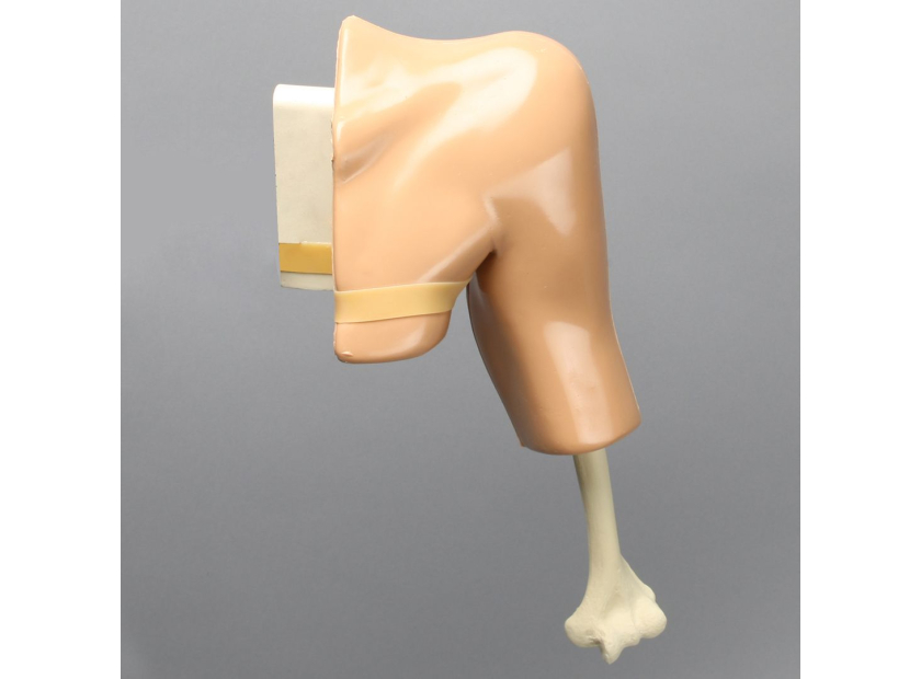
Labrum tears can involve both the labrum and the bicep and can happen anteriorly or posteriorly. Having a shoulder joint model with ligaments can help improve accurate diagnosing and surgical treatment for specific labral tears by giving students an authentic experience of inspecting the shoulder anatomy. The following advanced features of a shoulder joint model with ligaments are necessary to increase the efficiency of labrum repair training:
- Labrum and biceps tendon: Shoulder joint models equipped with both the labrum and biceps tendon allow for demonstration of localized labral tears as well as tears involving the bicep, which are seen more commonly in SLAP tears. Surgical residents will be able to see how the different tendons affect shoulder stability and how to best reattach them if necessary.
- Fully articulating humerus: An articulating bone model provides a similar range of kinetic motion to the joint it is demonstrating. Having a fully articulating humerus allows surgical residents to see how the upper arm bone, also known as the humeral head, rests and moves within the socket. This allows trainers to show the extent of motion of a healthy shoulder as well as the restricted movements caused by a labral tear.
- Removable red muscles: Being able to demonstrate the musculature of the shoulder joint model will give students better visualization of the glenoid and give them a better understanding of how the upper arm bone fits into the socket.
- Acromioclavicular (AC) joint ligaments: The acromioclavicular joint is the joint where the collarbone, or clavicle, and scapula, or shoulder blade, meet. These ligaments give students a broader view of the shoulder’s anatomy, which is especially helpful when training for labrum repair procedures and understanding where to make incisions and insert the arthroscope.
Choosing the best shoulder model for surgical demonstrations is essential to increasing the effectiveness of labrum repair training. Having a shoulder joint model with ligaments gives more realism to the training approach to help students better assess labral tears in patients, decide when surgical intervention is necessary, and sufficiently repair any variation of a labral tear. Having a more realistic shoulder joint model to train with repetitively improves surgical outcomes with reduced errors and operation times.
The demonstration models created by Sawbones are designed to increase the efficacy of your surgical training program. For more information on our offerings or to talk about custom training models, contact us at 206-463-5551.
If you're seeking something you can't find on our website, our sales team is happy to help. We can either direct you to the right model or provide a free quote on the right custom project to meet your needs. Discover options with our clear bone models, laminated blocks, custom displays, or other machining projects.

Work with the industry leader in medical training products!
For four decades, Sawbones has offered the best medical training products for doctors, educators, sales specialists and more. Contact us to learn more about our high-quality models!
1 (206) 463-5551Contact Us!


