
Osteoporotic Bone Models: Looking at Common Fractures
An estimated 10 million Americans above 50 years of age have osteoporosis, with around 1.5 million suffering osteoporotic fractures yearly. A high-quality osteoporotic bone model aids in instructing residents about common fractures and injuries due to bone fragility. Below are several examples.
Osteoporotic Bone Models
Osteoporotic bone models demonstrate loss of bone density and mass. Using models, instructors can effectively show how osteoporosis thins cortical bone and increases porosity in the spongy bone.
Below are common osteoporotic fractures that can be discussed using osteoporotic bone models:
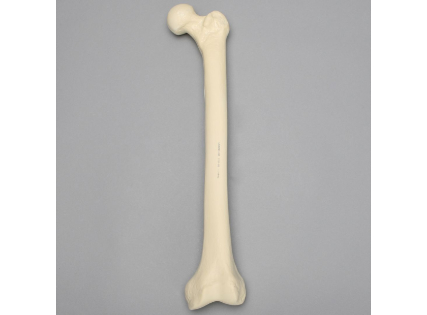
Femur
|
Femoral Neck Fractures |
Models show how the disease weakens the strength of the femoral neck and increases the risk of falls. Fractures of the femoral neck result in blood supply disruption, affecting healing, especially in elderly patients. Residents can discuss reduction and fixation techniques to restore mobility and function. Instructors can also use models to demonstrate modern techniques such as biologic bone augmentation to decrease the chances of fracture collapse following surgery. |
|
Closed Femur, Shaft |
Osteoporotic bone models assist residents in studying common spiral and transverse fractures of the shaft, as well as comminuted fractures. Residents can discuss closed reduction techniques to correct bone alignment. |
|
Open Femur, Shaft |
Open fractures of the femur shaft present a higher risk of complications due to skin, muscles, and tendon damage. Instructors can use osteoporotic bone models to discuss the open reduction and fixation techniques as well as best practices for minimizing damage to major ligaments and tendons. |
Forearm
|
Closed Distal Forearm |
Discussion can focus on isolated fractures in either ulna or radius, which have a higher chance of healing under non-operative management. Instruction can also emphasize how fractures in both the radius and ulna nearly always require surgical reduction and/or fixation. |
|
Open Distal Forearm |
Again, fractures in both bones indicate surgical intervention. Instructors can demonstrate how complex fracture patterns may necessitate repeat procedures due to the increased risk of complications. |
Humerus
|
Proximal and Distal Fractures |
Instructors can discuss which fractures are indicated for operative intervention, such as intercondylar and bicolumnar fractures. Models show how non-displaced fractures can be remedied using immobilization techniques. Osteoporotic bone models can also be used to discuss associated distal fracture injuries such as elbow dislocation. Proximal fracture discussion can focus on interventions ranging from sling immobilization to surgical fixation and arthroplasty based on the complexity of the injury. |
|
Shaft Fractures |
Models show different locations of shaft fracture, i.e. proximal, middle, or distal third. Instructors can discuss fracture patterns and recommended interventional techniques. Residents can practice non-operative techniques such as coaptation splints, functional braces, and damage control orthopaedics. In addition, models show which operative technique is best for shaft fractures based on injury location. For example, an anterior approach is used for ORIF procedures applied to middle third shaft fractures, while an anterolateral approach works best with proximal third to middle third fractures. |
Spine and Vertebrae
|
Pathologic Vertebral Fractures |
Models show how osteoporosis can narrow and flatten vertebrae, causing elderly patients to bend forward. Osteoporotic bone models demonstrate common characteristics of vertebral compression fractures such as decreased bone quantity, thinned cortices, and decreased cancellous bone trabecular continuity. Models also demonstrate the rationale of interventions such as bracing, vertebroplasty, kyphoplasty, and surgical decompression. |
|
Spine Fractures |
The spine is especially prone to osteoporotic fractures as it functions similar to a long bone like the femur or humerus. Osteoporotic bone models show how fracture can stem from spine immobility and resorption due to loss of bone density. The cervical spine is also susceptible to osteoporotic fractures due to its location at the base of a mobile head and neck while being fused to a thoracic area. Residents can discuss different interventions such as vest placement or surgical procedures, ex fixation techniques. |
|
Lumbar Fractures |
Osteoporotic bone models show how even low-energy stress and impact can cause lower spine fractures due to bone insufficiency. Instruction can dive into fracture patterns such as flexion, extension, and rotation. Residents can use bone models to study indications for surgery such as comminution, obvious reduction of vertebral body height, excessive angulation, and associated injuries such as ligament damage. |
|
Thoracic Fractures |
Again, osteoporotic bone models show how the disease contributes to compression fractures due to bone thinning. Bone insufficiency during relatively normal activity such as bending to pick an object from the floor or falling from standing height can cause thoracic compression fractures. Residents can use bone models to discuss non-operative techniques such as bracing or operative intervention, including fixation. |
Osteoporotic bone models help residents visualize key characteristics of osteoporotic fractures, such as increased fragility, loss of density, and thinning bone, among others. Bone models show common fractures to specific areas such as the femur, humerus, forearm, and spine, along with recommended interventions.
Models also help demonstrate how fractures can occur from relatively low-energy impact due to bone insufficiency. Lastly, osteoporotic bone models can be used to study surgery indications along with surgical approaches and techniques.
Best-In-Class Osteoporotic Bone Models
Sawbones offers a range of high-quality osteoporotic bone models, including osteoporotic femur, osteoporotic lumbar and thoracic vertebrae, cross-section osteoporotic vertebrae, and more. Our collection includes foam models, foam cortical with cancellous inner material, radiopaque models for medical imaging, and scanned bone models in standard CAD file formats.
Sawbones offers the best-in-class osteoporotic bone models for medical demonstration and instruction. For more information on our offerings or to talk about custom training models, contact us at 206-463-5551.
If you're seeking something you can't find on our website, our sales team is happy to help. We can either direct you to the right model or provide a free quote on the right custom project to meet your needs. Discover options with our clear bone models, laminated blocks, custom displays, or other machining projects.

Work with the industry leader in medical training products!
For four decades, Sawbones has offered the best medical training products for doctors, educators, sales specialists and more. Contact us to learn more about our high-quality models!
1 (206) 463-5551Contact Us!


