
How To Use Anatomical Skull Models For Trauma Training
Traumatic brain injury represented around 600+ related hospitalizations per day in 2019 and 176 associated deaths per day in 2020 in the US, based on statistics reported by the Centers for Disease Control and Prevention (CDC). Surgeons in training need to know how to evaluate skull fractures when examining common head injuries. A high-quality anatomical skull model helps residents build familiarity with trauma injuries and subsequent surgical procedures. Let's take a closer look.
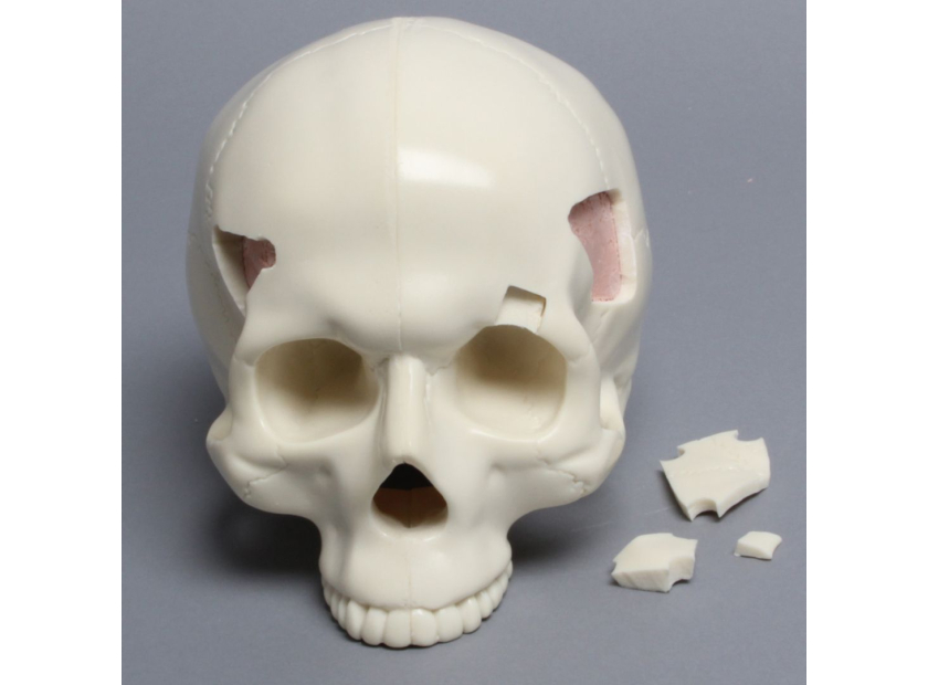
Anatomical Skull Model For Trauma Training
A lot of thought goes into decision-making whenever a head injury presents indications for surgery. Surgeons must always balance the risk of possible irreparable damage to the brain with the patient’s chances of survival. Such decisions often have to be made in a couple of hours, underlining the need for swift assessment and sound judgment of traumatic head injuries. Anatomical skull models help familiarize residents with common injury types as well as improve the following skills:
- Anatomical knowledge of the skull - Trainees gain detailed knowledge of the skull’s parts, structure, and anatomical functions
- Identification of common skull fractures - Models demonstrate the type of trauma injuries and resulting fractures, ex. linear fracture, depressed fracture, diastatic fracture, and basilar skull fracture
- Surgical outline and incision - Surgical incision landmarks and techniques can be practiced on anatomical skull models. Their relation to the injury as well as special areas of interest, ex. hematoma, hairline, etc., can also be discussed
- Skull fracture repair techniques - Various operative techniques for trauma injuries can be demonstrated in detail, along with their risks and advantages. Appropriate management of skull fractures concerning the brain, spinal cord, facial structure, neck, eyes, and other areas can also be rationalized during instruction using an anatomical skull model
- Fixation techniques - Options for bone fragment fixation can be demonstrated using anatomical skulls like inside-outside fixation, use of pins, clamps, cranial mini plates, etc.
- Cosmetic considerations - Correction of gross cosmetic deformities, minimization of scarring across the forehead, and operating behind the hairline are some cosmetic considerations to be discussed when repairing skull fractures
Open Skull Fractures
Assessment of skull fractures arising from traumatic injury is often tricky because neurological damage is sometimes absent for a period of time. In addition, the scalp may move from the site of the fracture and hide dural tears and other damage. Familiarity with open skull fractures can be emphasized during training in the following ways:
|
Indications for Surgery |
Anatomical skull models designed to display fractures can be used to emphasize indications for surgery, such as:
|
|
Skull Fracture Repair |
Techniques for repairing skull fractures can be demonstrated in detail, including:
|
|
Other Considerations |
Cosmetic considerations requiring bicoronal incision behind the hairline, etc. can be demonstrated using an anatomical skull model |
Decompressive Craniectomy
Another surgical intervention for head trauma and skull injury, decompressive craniectomy is intended to relieve pressure from the brain by diverting injured tissue through an opening. However, complications arise when possible risks and the actual efficacy of the procedure are considered. Anatomical skull models facilitate medical training and discussion of decompressive craniectomy in the following ways:
|
Indications for Surgery |
Given the lack of uniform indications for this procedure, anatomical skull models can demonstrate general guidelines, including:
|
|
Decompressive Craniectomy Techniques |
|
|
Other Considerations |
|
In summary, anatomical skull models facilitate instruction, demonstration, and discussion of trauma training for head and brain injury. Models provide visualization of common trauma types and resulting fractures to the skull. Anatomical models also demonstrate surgical techniques such as incision planning, repair procedures, and bone fragment fixation.
Lastly, anatomical skull models encourage discussion of special considerations in skull trauma surgery such as cosmesis, operative and non-operative management, preservation of dural venous sinus, contraindications, and more. All in all, residency programs level up the quality of instruction by using anatomical skull models for head and skull trauma training.
Improve Head Trauma Training With Sawbones Models
Sawbones manufactures anatomical skull models for various training courses. Our selection includes full skull models with and without vise attachment, skull models with soft-tissue and brain attachment, skull models with fracture and multiple fractures, skull caps with craniotomies, and more.
Sawbones offers the best-in-class anatomical skull model for medical demonstration and instruction. For more information on our offerings or to talk about custom training models, contact us at 206-463-5551.

If you're seeking something you can't find on our website, our sales team is happy to help. We can either direct you to the right model or provide a free quote on the right custom project to meet your needs. Discover options with our clear bone models, laminated blocks, custom displays, or other machining projects.

Work with the industry leader in medical training products!
For four decades, Sawbones has offered the best medical training products for doctors, educators, sales specialists and more. Contact us to learn more about our high-quality models!
1 (206) 463-5551Contact Us!


