
Rotator Cuff Repair: Enhanced Learning Through Anatomy Model Training
Age places patients at increased risk of suffering a rotator cuff injury. 50% of patients aged 66 and above are likely to suffer a bilateral rotator cuff tear, while an estimated 62% of patients 80 years old and above suffer from a range of rotator cuff injuries. When training for various rotator cuff repair procedures, residents can benefit from anatomical models. Let’s take a closer look.
Rotator Cuff Repair
Despite breakthroughs in rotator cuff surgical techniques, incomplete healing, and injury recurrence remain major concerns. Anatomical familiarity and precise surgical techniques are essential in increasing patients’ chances of recovering their original range of motion.
Anatomical models help familiarize residents with rotator cuff repair procedures. High-quality models also permit repetitive and hands-on practice to improve surgical dexterity with small arthroscopic instruments.
Hands-On Training
Rotator cuff repair procedures involve using arthroscopic procedures and equipment. Anatomical models sustain the necessary repetitive practice in order to master the following psychomotor skills:
Small Hand Movements
An arthroscopy requires precise small hand movements inside restricted spaces such as the shoulder, knee, and foot. Anatomical models aid residents in first visualizing the shoulder area, and then maneuvering around the rotator cuff and its parts. Models allow residents to progress at their own pace and gradually build psychomotor skills compared to cadavers, which only allow limited opportunities for practice.
Small Instrument Handling
In the same way, anatomical models help residents to become acquainted with specialized arthroscopy tools such as the arthroscope, shaver, and graft spreader, among others. Models aid surgical trainees in mastering instruments and building efficiency in procedural skill, helping them to complete surgical procedures confidently and quickly.
Techniques
Anatomical models and shoulder assembly sets assist residents in mastering rotator cuff repair techniques including:
- Diagnostic Arthroscopy
Models that aid in visualizing the shoulder and its anatomical parts. Residents can also use models to practice portal placement and tissue debridement prior to a major rotator cuff repair procedure.
- Bursoscopy
Using the model, trainees practice handling the obturator and maneuvering among the deltoid, acromion, and bursal curtain. Instructors can use models to demonstrate proper positioning of the scope to achieve maximum visualization of the bursal space.
- Anchor Placement
Best practices for anchor placement can be demonstrated using models. Techniques include using separate skin punctures for individual anchor placement, avoiding tension on the repair by precisely positioning sutures, and using additional anchors for large rotator cuff tears.
- Allograft Reinforcement
Anatomical models aid residents practice allograft reinforcement techniques for rotator cuff repair. These include proper graft orientation, the introduction of the graft through the cannula, and graft placement.
Small hand motions such as rolling and unfolding the graft, suturing, and knot tying can be demonstrated on models for greater visualization. Dedicated anatomical models with foam and other suturable materials facilitate the best hands-on training for these techniques. In addition, residents can practice handling arthroscopic instruments such as graspers, knot pushers, and suture measuring devices on anatomical models.
Lastly, models also visualize the pros and cons of rotator cuff repair techniques. Instructors can use models to show how a graft improves mobility, especially in cases of large tears or fully torn rotator cuffs. Discussion of biological and mechanical scaffolds in rotator cuff repair procedures can also be facilitated using anatomical models.
- Wound Closure
Instructors can use models to demonstrate superficial and deep wound closure techniques. They can also demonstrate post-operative care, such as using prefitted slings and braces on a model to show how they reduce pressure on the rotator cuff repair after surgery.
Anatomical models help residents to practice, refine, and master rotator cuff repair procedures through hands-on training. Models also provide greater visualization and facilitate discussion of proper techniques and best practices. Residents can perform repeated practice procedures on models to improve surgical techniques. Lastly, surgical trainees can familiarize themselves with arthroscopic instruments involved in rotator cuff repair procedures using models.
Best-In-Class Anatomical Models For Rotator Cuff Repair Training
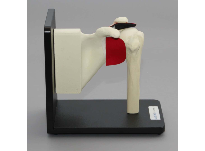
Sawbones offers specialized shoulder assembly sets and anatomical models for rotator cuff repair training. Our shoulder assembly with a rotator cuff on the stand comes with a neoprene rotator cuff.
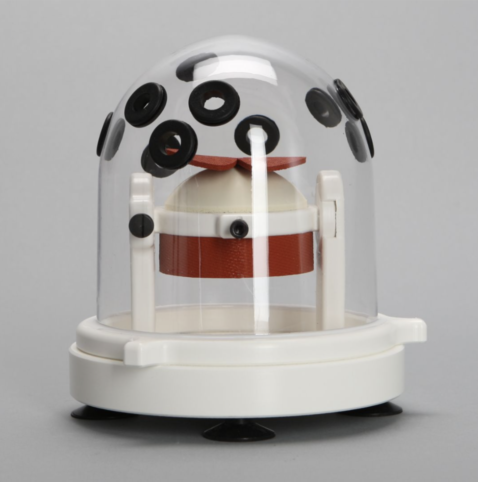
Dome Holder for Rotator Cuff and Labrum Repair
We also offer a shoulder assembly set on a stand with replaceable parts, including scapula, proximal humerus, rotator cuff, and supraspinatus ligament. This model comes with compatible screws and anchors.
Shoulder Assembly on Stand with Replaceable Parts, Foam Cortical

Sawbones, offers the best-in-class shoulder assembly anatomical model for rotator cuff repair training. For more information on our offerings or to talk about custom training models, contact us at 206-463-5551.

Work with the industry leader in medical training products!
For four decades, Sawbones has offered the best medical training products for doctors, educators, sales specialists and more. Contact us to learn more about our high-quality models!
1 (206) 463-5551Contact Us!


