
Spinal Vertebrae Models Must Contain These Features
There's a reason that the backbone is a metaphor for an object of support. These 24 vertebrae are responsible for both bodily function and protection of the nervous system. Regardless of specialty, any medical professional will need an in-depth understanding of it at some point in their career. That's why spinal vertebrae models are the backbone of a good education.
Necessary anatomical features will vary widely by course of study. For some, understanding the connection between muscle, ligaments, and bone will be as far as their education goes. For others, they'll need a working model that allows them to practice high-risk medical procedures in a realistic environment.
Necessary Anatomical Features of Spinal Vertebrae Models
The necessary anatomical features of spinal vertebrae models go far beyond the basic cervical, thoracic and lumbar regions. Most medical professions will deal with the dysfunction of systems localized in one of these spots. Standard anatomical features useful in medical training include:
- Intervertebral discs – Up to 2% of the adult population will suffer a herniated disc annually. While that figure doesn't seem like a lot, put into context, that's about 6.5 million people per year in the US alone. That makes this a widespread condition anyone in an orthopedic or neuropathic specialty will eventually treat.
- Psoas muscle – Extending from the spine to the femur, this muscle is the primary connection between the torso and lower limbs. Its continued function is necessary for virtually all activities, including sitting, walking, running, and dancing. This muscle is a significant focus for individuals working in specialties like chiropractic, physical therapy, and occupational therapy. A thorough understanding of the function of this muscle, as well as release techniques, is vital.
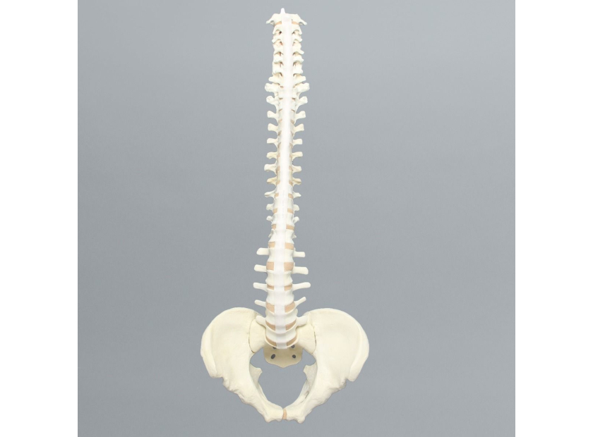
- Sacrum – The sacrum is at the base of the lumbar spine and is responsible for pelvic stability. The thing to remember when choosing a model for this is gender. A male sacrum is much different from a female one for obvious reasons. As women have shorter, wider sacra, they're more likely to suffer pain and injuries there.
- Scoliosis – Scoliosis isn't an anatomical feature. It's a spinal curvature that leads to dysfunction. In children, this condition is correctable with back braces that take advantage of the bone's malleability during youth. In adults, surgery is often advisable in severe cases. Models that display this condition and realistic ones that allow practice procedures are crucial for many specialties.
- Cancellous bone tissue – This spongy bone tissue is prevalent throughout the spine, particularly at the core of vertebrae bones. It's a rich source of osteoprogenitor cells needed in the formation of new bone. As such, it's the useful source material for bone grafts required in spinal fusion surgeries and other complicated procedures. Both surgeons and those who harvest these materials at bone banks need a thorough understanding of removal methods that preserve integrity.
- Anterior and posterior longitudinal ligaments – These important ligaments have varying thicknesses, textures, and connections to vertebral bodies requiring in-depth examination. The ability to review these ligaments' superficial, intermediate, and deep layers will depend on the spine model’s detail.
The anatomical features of the spine are virtually endless. The above incorporates a few of the more prominent areas likely to come into play during medical training. Regardless of the model's features, some material considerations are essential to ensure a well-rounded medical curriculum.
Material Considerations in Spine Models
There are many material considerations in orthopaedic models to consider. When it comes to medical training, it's always best to opt on the side of realism, both in aesthetic and texture. Solid foam materials are typically the most economical when it comes to teaching general anatomy. However, there are a few additional features to consider for more in-depth lessons, specifically when dealing with the spine.
One of the most prevalent is whether radiopaque properties will be necessary. Teaching students how to diagnose fractures in the spine and sacrum—which are incredibly complex—is one area where this is useful. Another lies in scoliosis diagnosis, which is usually verified via MRIs, CT scans, or radiographs.
The model's flexibility and visibility are also considerations. Reinforced flex rods allow instructors to bend and rotate the model to demonstrate different movements and functions. Windows within it may offer a view of the inner materials in evaluating bones, soft tissues, disc nuclei, and compressed nerves.
In many cases, the spinal vertebrae model will have to be sourced for a specific purpose, whether diagnosing a condition, practicing a surgical technique, or in medical imaging training. Instructors should review the lesson plan thoroughly to find the model that encompasses all their various requirements. When in doubt, it's best to err on the side of caution by choosing the most detailed model.
Sawbones has an extensive catalog of spinal vertebrae models for everything from anatomical instruction to surgical practice. To learn more, contact us at 206-463-5551.

If you're seeking something you can't find on our website, our sales team is happy to help. We can either direct you to the right model or provide a free quote on the right custom project to meet your needs. Discover options with our clear bone models, laminated blocks, custom displays, or other machining projects.

Work with the industry leader in medical training products!
For four decades, Sawbones has offered the best medical training products for doctors, educators, sales specialists and more. Contact us to learn more about our high-quality models!
1 (206) 463-5551Contact Us!


