
Skull Bone Structure: Tips for Using Cranial Models for Demonstration and Teaching
Medical students learn more effectively through demonstration than description. That’s just the price of having to assimilate complex procedures and tool use into a working repertoire of specialized skills. The challenge for instructors is finding resources and models that can reliably instill these potentially life-saving skills.
That’s where the best-engineered teaching and training models become crucial. Using an advanced skull bone structure model, for example, takes learning a range of cranial procedures and diagnostic knowledge beyond the simple retention and recollection of facts. Realistic materials, interactive features, and layer upon layer of detail help deliver skills that can be transferred directly to practice for the benefit of real-world patients.
From hands-on psychomotor skill trainers, such as skull models with endoscope capacity, to highly detailed models that demonstrate the core skills of craniotomy, using the most advanced training resources makes medical instruction more effective.
Demonstration Over Description
Research based on years of evolving medical instruction has found that teaching the skills needed for a specialist vocation is different than teaching basic content. Simple 2-,3-, and 5-step procedures can be effectively taught using Socratic lecture principles, but the far more complex procedures that must be mastered by medical professionals require a more visual and hands-on approach. To get to the finer detail of diagnosis, intervention, and even core clinical skills such as patient examination and synthesizing data, you need more realistic teaching tools.
The use of advanced models gives trainees the opportunity for repeated practice with immediate feedback so they can refine their skills without impacting patient safety.
The advantages of a model-led teaching approach are clearly evident in the features of cranial simulations that can be used to teach skull bone structure across a range of disciplines.
Features of Advanced Training Models
The interactive training models available to today’s medical instructors are highly specialized teaching tools that utilize advanced features. By combining the elements below, instructors can create demonstrations and hands-on practical engagements that faithfully reconstruct the presentations and procedures specialists will encounter in the field.
These features include:
- Haptic Feedback: Models are equipped with sensors that emit sounds upon contact. This form of instant response creates repercussions for inaccurate movements and guides the development of psychomotor skills and triangulation.
- Composite Bone: The human skull is not made of uniform bone. Today’s most effective models authentically reproduce variations in the frontal, parietal, sphenoid, and temporal bones, and the contours of the mandible.
- Radiopaque: The density of this material helps block radiation, making it useful for medical imaging training.
- Dual Usage Models: Students can train on open and closed variations of the same model to aid in arthroscopic and craniotomy techniques.
- Buildable Models: Advanced models include variations that can evolve with student competencies. This dynamic potential gives students the time to learn through repetition, and the learning curve to reflect the fluid nature of ever-evolving best practices.
Incorporating these dynamic features within the specific anatomy and procedures of the skull results in an impressive range of teaching tools. Perhaps more importantly, these models can be augmented with custom pieces to round out a specialist teaching module for novices or professionals pursuing new training.
Skull Bone Structure Models for Every Purpose
The features above, and many others, can be combined to create specialist teaching aids that provide the dynamic learning environment proven to improve learning outcomes. Here are some examples of how these combinations can be stretched to fill almost any requirement.
Skull Model for Endoscope Training
A supine model with facial muscles and complete sinus, this training aide is accessible by endoscope, giving students a reusable platform for mastering the triangulation and psychomotor skills of the device. Endoscopy has become one of the most important diagnostic and surgical tools of the modern era with its low-risk profile and minimal patient impact, but it takes many hours of training to master its full potential.
Skull with Fracture and Craniotomies
This model has cranial flaps on both sides of the skull and soft tissue brain matter to simulate the demands of open craniotomy. It provides the instructor with a dynamic demonstration of the less invasive craniotomy burr technique for interventions against subdural hematoma and brain tumors.
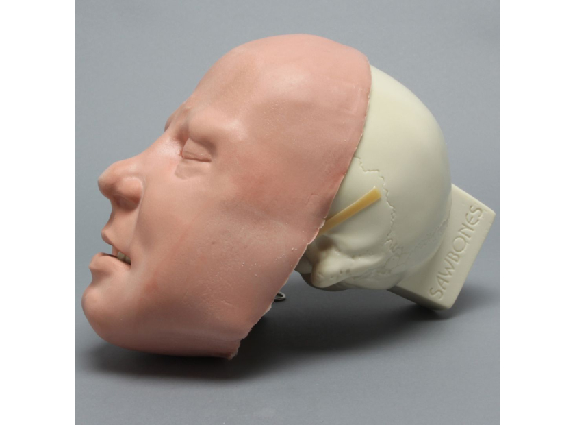
Skull with Fractures and Soft Tissue Face
This full skull model includes predrilled holes for wiring between teeth, cheekbone fractures, working mandible, and a full soft tissue face for increased performance. It’s an authentic, reusable upgrade from the expense and handling of a cadaveric model, and a safe alternative to clinical presentation.
These models, and others like them that simulate a range of specializations from genioplasty and pediatric to veterinary interventions, represent the next generation of teaching tools that offer the key benefits of affordability and reusability that are ushering in an era of greater standardization of medical competencies.
Standardization for Better Healthcare
Standardization of medical training, in both the novice and professional development modes, can lead directly to increased patient safety, continuing quality of care improvements, and the more effective spread of best practices.
Advanced training models such as the skull bone structure examples above are a key resource in attaining greater standardization, at least within specialities. These models are affordable and accessible, they are reusable and repeatable, and they are proving effective.
Crucially, the continuing partnership of leading medical educators with advanced model engineers means that highly specialized teaching tools can be created rapidly and to commercial scale to disseminate advances in diagnosis, intervention, and education itself.
Skull Bone Structure Models that Better Engage
The key to unlocking this potential for standardizing medical instruction across any given field is creating models that engage and inspire students. The complex nature of medical training means models such as those used to teach the intricacies of skull bone structure need to be three-dimension, authentic, and hands-on. Medical procedures extend well beyond the 5-step limits of descriptive learning and instead need additional vigor.
The next generation of medical students needs the next generation of dynamic educational models.
The educational models created by Sawbones are designed to enhance the impact of your lessons. Our engineers can work with you to create unique, specialized solutions that transform your knowledge into teachable engagements. Contact us today online or call us at 206-463-5551 to learn more.
If you're seeking something you can't find on our website, our sales team is happy to help. We can either direct you to the right model or provide a free quote on the right custom project to meet your needs. Discover options with our clear bone models, laminated blocks, custom displays, or other machining projects.

Work with the industry leader in medical training products!
For four decades, Sawbones has offered the best medical training products for doctors, educators, sales specialists and more. Contact us to learn more about our high-quality models!
1 (206) 463-5551Contact Us!


