
Using Foot and Ankle Anatomical Models for 5 Diagnostic and Operative Arthroscopy Techniques
Feet contain ¼ of all the bones in the body (52 out of 206) in a relatively small space compared to the rest of the body. Their complex structure presents a challenge to ordinary surgical procedures. Typical imagery methods, including X-rays, MRI, and CT scans also do not detect injuries in the foot and ankle ligaments and tendons. Arthroscopy is a better way to identify and treat the causes of foot and ankle pain.
Accurate foot and ankle anatomical models help orthopaedic trainees build diagnostic and operative skills used in arthroscopy. Let’s take a closer look below.
Uses of Foot and Ankle Anatomical Models
1. Foot and Ankle Anatomy
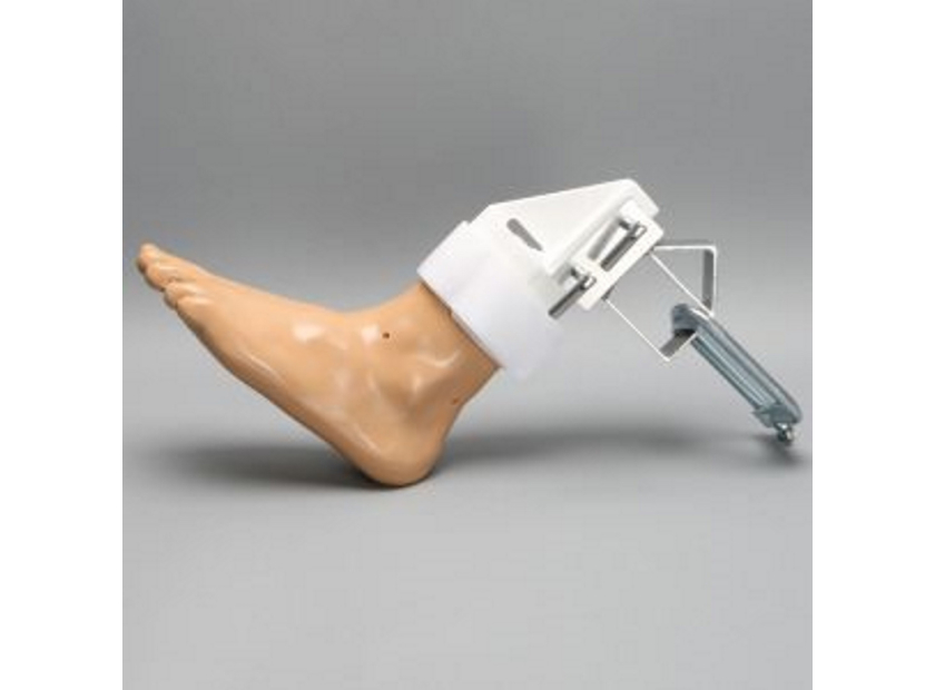
The success of arthroscopy procedures lies in thorough familiarization with foot and ankle anatomy. In addition to numerous bones, the foot has 33 joints, 19 muscles, 107 ligaments, and several tendons in an already tight space. The intricate anatomy of the foot and ankle demands great skill and extraordinary care to avoid damage to major arteries and veins. High-quality models help medical students and orthopaedic trainees master anatomy for visualisation and operative techniques.
In a study with 48 orthopaedic residents from across the United States and Canada, researchers found that training with a simulator decreased camera probe distance and time to completion. These results represented significant improvements in residents’ familiarization with anatomy as well as instrument handling after training with a simulator for arthroscopy.
2. Portal Placement
Foot and ankle anatomical models help orthopaedic trainees practice portal placement in a risk-free environment. Indeed, another study published in ResearchGate determined that the use of low-fidelity models in arthroscopic simulation training improved surgery skills in basic ankle arthroscopy. Models help illustrate which nerves and veins are at risk from specific techniques, such as:
- Anterocentral - high risk to dorsalis pedis artery (dorsal artery of foot) and peroneal nerve
- Anteromedial - endangers saphenous nerve, risks injury to tibialis anterior tendon
- Posteromedial - endangers posterior tibial artery
Anatomical models demonstrate the merits of each technique as well as their risk of complications. Models also highlight techniques that require a higher degree of surgical skill to perform.
3. Surgical Instruments
Arthroscopy necessitates small diameter instruments in tight anatomical spaces such as the foot and ankle. Operative techniques, therefore, demand dexterity with small joint instruments, including shavers, knives, abraders, curettes, and others.
In foot and ankle arthroscopy, safe usage of a high volume fluid system (and possibly infusion pump) is critical. Foot and ankle anatomical models help trainees become familiar with surgical instruments and equipment for a successful operation.
4. Arthroscopy Techniques
The following foot and ankle arthroscopy techniques can be practiced with anatomical models, such as:
|
A) Diagnosis and Visualisation |
|
|
B) Manoeuvre of Arthroscopic Device |
|
|
C) Anterior Portal Placement |
|
|
D) Anteromedial Inferior Portal Placement |
|
|
E) Posterior Coaxial Portal Placement |
|
5. Medical Training and Patient Education
Medical professionals other than orthopaedic surgeons also profit from practicing with high quality anatomical models. Chris Watford, a graduate of Medical Sales College, shared that his knowledge of foot and ankle procedures ultimately helped him land a medical sales job. He said, "During my interview I gave a presentation similar to the presentations we trained for and practiced during the program. The presentation was for the five most common surgical foot and ankle procedures."
Foot and ankle anatomical models also improve patient education. Surgeons can use models to show how arthroscopy procedures are designed to be minimally invasive yet effective treatments for foot and ankle problems.
Best-in-Class Foot and Ankle Anatomical Models
In summary, foot and ankle anatomical models help orthopaedic trainees practice diagnostic and operative techniques in arthroscopy. High quality models demonstrate the merits and risks of different techniques, particularly the danger of neurovascular injury. Models also help medical students become familiar with small diameter tools and equipment necessary for successful foot and ankle arthroscopy. Lastly, foot and ankle anatomical models improve patient education and understanding of arthroscopy procedures.
Sawbones offers best-in-class foot and anatomical ankle models for orthopaedic training and medical education. Sawbones is known for originating hands-on workshop models and continues its leadership today in manufacturing anatomical medical training models. For more information on our offerings or to talk about custom training models, contact us at 206-463-5551.

If you're seeking something you can't find on our website, our sales team is happy to help. We can either direct you to the right model or provide a free quote on the right custom project to meet your needs. Discover options with our clear bone models, laminated blocks, custom displays, or other machining projects.

Work with the industry leader in medical training products!
For four decades, Sawbones has offered the best medical training products for doctors, educators, sales specialists and more. Contact us to learn more about our high-quality models!
1 (206) 463-5551Contact Us!


