
Dynamic Features of 8 Lumbar Spine Models For In-Depth Medical Training
The lumbar vertebrae are the hinge of the body. From L1 to L5 pivot the major muscles of the back and the nerve roots of the spinal cord that both drive movement and form the body’s center of gravity. The torque and weight forces that are constantly in operation throughout the area leave it prone to a range of injuries, and its tightly packed anatomy means the resulting pain can radiate well beyond the source.
Successful interventions associated with herniated or degenerated discs, compressed nerve roots, muscle spasm and strain, and spinal deformities have to be undertaken with strict reference to the entire physiology. In response to the complexity of these interventions, dozens of highly specialized lumbar spine models have been developed that use dynamic features to train practitioners.
Eight of the best examples of these next-generation models are listed below, and more dynamic and responsive models are on the way as instructors and engineers collaborate to produce the most realistic and effective training aides yet.
The Essentials of Medical Training Models
Medical training models have a clear appeal in all branches of specialist education—they provide an affordable, accessible, and safe alternative to the use of cadavers or reliance on supervised operating room exposure.
Engineered models can be custom-made as a resource in the teaching of highly specialized procedures and can encourage a professional level of standardized best practice among students and experts alike.
To successfully transfer procedural knowledge and fundamental surgical skills to the operating room, the current generation of models have several dynamic features that better replicate real world conditions than any previously available:
- Haptic Feedback: Models produce a noise when instruments contact internal elements. This instant response encourages greater accuracy and efficiency and introduces repercussions to errors.
- Buildability: Training models can be expanded to increase difficulty and introduce new presentations as students progress.
- Composite Bone: This is a realistic reproduction of the strength, feel, and vulnerabilities of bone. Dramatic improvements have also been made in the replication of skin, muscle, and tissues to improve instrument use.
- Open and Closed Models: Models can be altered to mimic open surgical interventions or closed arthroscopic procedures.
- Radiopaque: Materials block radiation, making it useful for medical imaging training.
- Solid Foam and Cortical Shells: Models are made of material robust enough to recreate cutting and drilling procedures and to withstand use across cohorts to encourage standardized best practice.
These dynamic features, among many others, can be combined to create highly specialized models that help reduce the learning curve of trainees.
8 Dynamic Lumbar Spine Models
Variety and specialization are demonstrated in the most advanced lumbar spine models. More importantly, leading model engineers are able to produce customer resources in collaboration with medical instructors to further improve medical training.
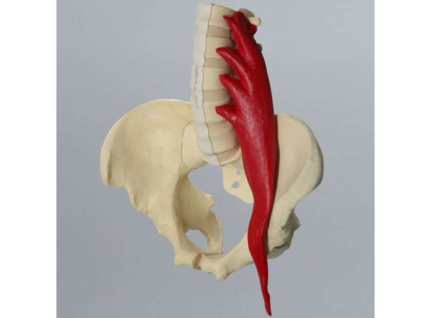
1. Lumbar Spine with Full Pelvis and Psoas Muscle
One of the most economical options available, this solid foam model of the lumbar and pelvis includes the psoas muscle, one of the common sites of lower back pain. Tightness of the muscle can compress lumbar discs, and inflamed psoas can impact the ilioinguinal and the iliohypogastric nerves.
2. Lumbar Spine with Full Pelvis and Nerve Roots
This male pelvis model includes nerve roots and dura and gives a neurosurgeon’s overview of the area. Semi-rigid discs allow for limited movement and clearly define the complexities of the lower back. The model can be an aid in diagnosing the nerves responsible for degenerative disease.
3. Radiopaque Lumbar Spine
As mentioned earlier, radiopaque models are designed for medical imaging training and block radiation, like that used in x-rays. In the case of lumbar models, the goal is to provide realistic imaging so students understand how to diagnose problems associated with the area.
4. Spine, Scoliosis
An authentic recreation of thoracic-lumbar scoliosis, this model features flexible discs and a malleable rod for limited fixation and manipulation. The model can be a valuable tool for understanding the complexities of spinal fusion surgery. The new generations of medical training models are capable of accommodating hooks, screws, and other methods of connecting two or more bones in the spine.
5. Lumbar Vertebrae with Nucleus Filled Discs
Featuring an extractable representation of the soft filling at the center of intervertebral discs, this model gives students a practical guide to the treatment of herniation and degeneration.
6. Dura Repair Kit
The perfect training kit for repairing the dural tears that occur during surgery, this model provides practical education in one of the fundamentals of surgical practice. The kit includes a gravity drip line with fittings and can be reused to hone delicate motor skills.
7. Spine Lumbar, Nerves, Muscles, and Cartilage
One of the most detailed lumbar spine models available, this unit gives students complete access to the intricacies of the region. Colorfully designed and built from the latest in composite materials for authentic response, this model can become a cornerstone of lumbar instruction.
8. Radiopaque Lumbar with Nerve Roots, Flava, and Interspinous Ligaments
A fine example of how the dynamic features of modern medical resources can be combined to increase specialization, this model offers both anatomical and diagnostic advantages. The composite materials are radiopaque to encourage the use of medical imaging, and the structure is dynamic enough to withstand practical manipulation.
As with all the advanced lumbar models detailed above, this model was designed to give a broad assessment of the lumbar region and facilitate training in a specific procedure or range of interventions.
The advantage of modern model development is that pieces such as these can be the starting point for customized tools that combine the dynamic potential of engineered design toward a highly specialized outcome.
Create Your Own Lumbar Spine Model
The current generation of medical model engineers is creating products, displays, and training solutions that maximize the knowledge of educators. By combining the latest in material and response technologies with cutting-edge teaching practices, you can make your own training tools. These tools can be specifically delivered just for your purposes, or commercially produced to aid standardization of best practices.
The modern answer to the challenges of preparing specialists for careers working in highly complex areas such as the lumbar spine is to refine and enhance teaching tools toward more realistic and effective outcomes.
As a creator of innovative medical training models, Sawbones is your partner in developing solutions that demystify the complexities of best practices. Our engineers can produce models to your specifications that make use of the most dynamic teaching technologies of performance, feel, and durability. Contact us today online or call us at 206-463-5551 to learn more.

If you're seeking something you can't find on our website, our sales team is happy to help. We can either direct you to the right model or provide a free quote on the right custom project to meet your needs. Discover options with our clear bone models, laminated blocks, custom displays, or other machining projects.

Work with the industry leader in medical training products!
For four decades, Sawbones has offered the best medical training products for doctors, educators, sales specialists and more. Contact us to learn more about our high-quality models!
1 (206) 463-5551Contact Us!


