
Using Human Torso Anatomy Models in Medical Training
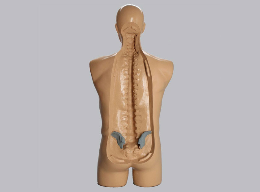
Human torso anatomy models are found in almost all forms of medical training. They are a lynchpin of the educational environment because they offer practical experience when materials are in short supply. Often, the only other option is human cadavers, and those are extremely difficult to obtain and certainly not for non-specialists.
Many procedures in the torso require repeated practice because precision is vital. Working around the spine is a high risk, so repeated practical training is imperative. By choosing the right human torso anatomy models, educational leaders can set their trainees up for success.
Human Torso Anatomy Models by Specialty
Human torso anatomy models have many applications in medical training. There are a variety of fields where these tools are indispensable.
Orthopaedics |
Pediatrics |
Chiropractic |
Physiatry |
| Artificial disc replacements, spinal fusions, discectomies, and treatment of degenerative afflictions are significant parts of this practice. Models are needed for diagnosing conditions and recommending treatment plans. | Childhood maladies like scoliosis and discitis require extensive study to ensure pediatricians can accurately diagnose and treat these issues. These models must help them understand how the problem will change as the child grows. | Spinal column misalignment correction is the majority of most chiropractic work. Accurate models help them understand and correct disorders and practice standard options like the Gonstead technique or flexion distraction. | Helping patients restore function and overcome disabilities often centers on treatments to the spine and torso, especially following surgical care. Chronic back pain management is often an area of specialty for these students. |
Neurosurgery |
Pain Management |
Rheumatology |
Physical Therapy |
| Neurosurgeons require an extensive understanding of the spine and its impact on the spinal cord. Models help them understand everything from degenerative issues to congenital defects like spina bifida. They will also help them practice a myriad of surgeries to correct such issues. | The administration of pain control injections and blockers is a highly delicate process. Doctors will need extensive practice before giving facet injections or cortisone shots. Spinal cord stimulation is another area where detailed training is vital. | Diseases of the joints, bones, and muscles are common in the torso. Rheumatologists need to experience diagnosing and treating these conditions and how they lead to issues like spinal stenosis and disc herniations. | Physical therapy is often the first stop after a traumatic back injury. Physical therapists need training in heat therapy, mobilization, massage, and exercises to strengthen and stabilize the spine. |
Torso models are necessary for medical training, especially when the only alternative is hard to obtain cadavers. Choosing the best possible model will help students gain the practical experience needed to secure their chosen specialty’s success.
Tips for Selecting the Best Anatomy Model
There are many options for human torso anatomy models, though not all are suited to medical training. When seeking out the best option, the following features provide the most comprehensive learning experience.
- Cosmetic realism: Medical training models are different from patient education displays. Patient models usually leverage highly contrasting, unnatural colors inappropriate in an educational environment. Colors and materials – especially markers use to show degeneration, disc material, vertebral arteries, and more – should appear as realistic as possible to provide the challenge students need for success.
- Patient-specific: Consider the specialty before choosing the torso model. For example, using an adult model for training on scoliosis treatment might be unrealistic, as treatment typically occurs in pediatric cases. A scoliotic pediatric model would be the better choice.
- Radiopaque properties: Radiopaque materials are materials that block radiation, like the kind found in x-rays. They are designed to show up under x-ray and fluoroscopy imaging. Extensive imaging is used to diagnose conditions in the torso, especially in the cervical and lumbar regions. Radiopaque models provide realistic imaging, so students understand how to identify these problems.
- Kinetic accuracy: The human torso is responsible for many of the motions and flexions in the body. The realistic kinetic movement teaches chiropractors, physical therapists, and occupational specialists how injuries occur and techniques to resolve issues.
- Buildability: The human torso is an extensive category that incorporates almost all the major organ groups and systems. It may be more cost-effective to start with a shell model and then build upon it as the course's complexity grows. Teachers gain access to the models they need and can expand on them as required.
The best human torso anatomy models for medical training will cater to specific audiences. Surgical type models will focus on anatomical fidelity and a true to life feel. In other training instances, the accuracy of motion may be more critical. The best person to select the type of medical model needed is usually designing the training curriculum. They'll know precisely what their students need to prepare for their future in medicine.
Sawbones offers a wide range of human torso anatomy models suited for many specialties. For more information on our offerings or to talk about custom training models, contact us at 206-463-5551.

If you're seeking something you can't find on our website, our sales team is happy to help. We can either direct you to the right model or provide a free quote on the right custom project to meet your needs. Discover options with our clear bone models, laminated blocks, custom displays, or other machining projects.

Work with the industry leader in medical training products!
For four decades, Sawbones has offered the best medical training products for doctors, educators, sales specialists and more. Contact us to learn more about our high-quality models!
1 (206) 463-5551Contact Us!


