
Skull Muscle Models: 5 Requirements for More Teaching Scenarios
The muscles of the skull are more challenging to a surgeon’s technique and knowledge than those of the limbs and torso. They are more complex in their innervation, slower in their fiber type, and have unique morphology that allows them to perform a broad range of intricate roles in chewing, movement, and facial expressions.
It takes a skilled hand to navigate this delicately tangled system in order to perform any number of interventions across the skull, from endonasal diagnosis to craniotomy.
Educating trainees on how to deliver successful surgical outcomes across this terrain requires skull muscle models with 5 key elements:
- Authentic composition and haptic response
- Buildability to increase model complexity
- Dual usage for arthroscopic and open simulations
- Durability for repeated use
- Customization to ensure accuracy
The right skull muscle models can reduce the learning curve for novice surgeons and afford them the repetition, standardized learnings, and authentic settings necessary to achieve the best possible patient outcomes.
The Importance of Models and Simulation Training
According to the Society for Simulation in Healthcare, models such as those that replicate the muscle and bone structure of the skull are an essential bridge between classroom learning and real-life clinical experience. The models are affordable, reusable, and scalable tools that offer trainees precious procedural exposure without risking patient safety or compromising limited cadaveric resources.
Johns Hopkins has developed an entire simulation center in Baltimore to expose surgeons to a wide range of variable and wholly customizable presentations within the broader context of a working hospital department. While the average medical teaching facility may not run to that extreme, the reported success of the Johns Hopkins approach validates the use of simulations such as skull muscle models and the tactile experience it affords novice professionals.
The transferable nature of the skills acquired through simulated surgical environments and presentations is related directly to the properties of the models in use. That’s why it is important that the next generation of teaching tools take advantage of modern engineering techniques to produce models designed to produce experts in any field or intervention.
1. Authentic Composition and Haptic Response
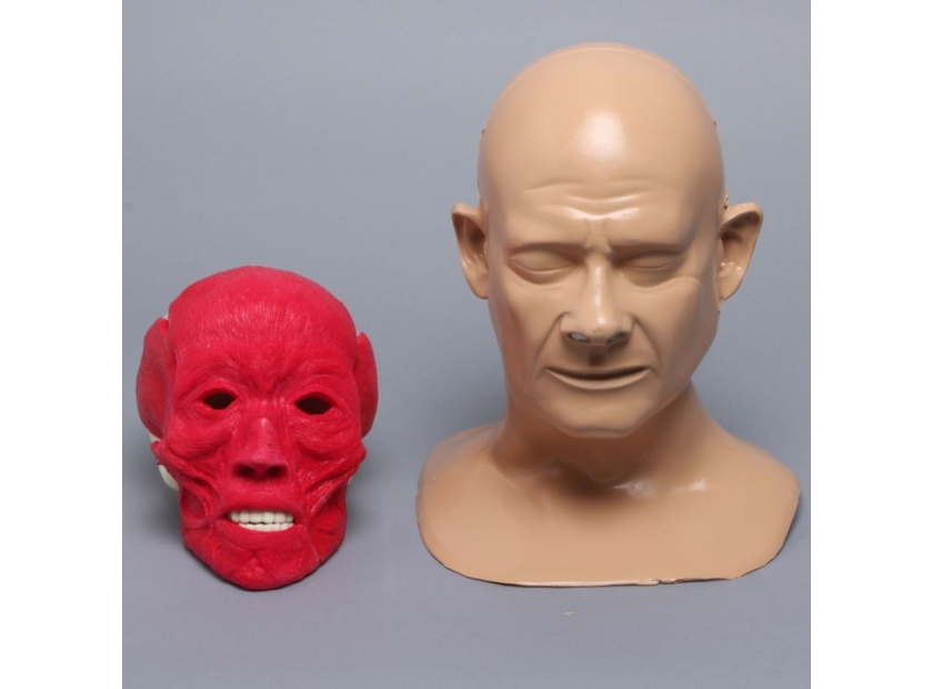
Cranial interventions require the use of a range of surgical tools against a spectrum of anatomical materials. The dense bone of the skull and the highly sensitive, innervated muscles of the face pose a unique challenge to inexperienced physicians. That’s why skull muscle models and training simulations must authentically recreate the resistance and performance of bodily structures.
Advances in bone composition and muscle modeling make today’s engineered materials, whether full or partial representation—complete with removable skin—an authentic replication.
Authenticity can be further improved with the use of haptic response technology that introduces consequences; through sound or vibration, haptic techniques add an extra level of challenge to a non-clinical setting.
2. Buildability to Increase Model Complexity
Surgeons learn through repetition—research has found it takes hundreds of procedures to master basic techniques—but there’s still a need to increase complexity in order to stimulate development. Model buildability gives teachers the power to introduce new variables and increased time, resources, or skill demands to continually progress students.
Engineered model solutions can be built to follow the development of specific skills for the completion of common procedures, or to hone the motor skills and triangulation challenges of using particular surgical tools.
3. Dual Usage for Arthroscopic and Open Simulations
Procedures involving the skull center around two main interventions—closed arthroscopic procedures and open craniotomy. Both have their advantages and are essential elements of surgical intervention involving the skull.
Skull muscle models can be engineered to offer practical experience in both approaches. Taking their cue from the instructor’s goals, models can even be created to progress from clear plastic to portal navigation and on to radiopaque features for medical imaging training.
4. Durability for Repeated Use
Teaching environments are testing grounds for surgeons. As such, the tools that shape their education and provide them with their first non-clinical surgical experiences need to survive the twin tests of longevity and relevance. Simulation models need to be robust enough to survive the surgical infancy of repeated cohorts. Models also need to be affordable enough to be replaced over time as advances in clinical and procedural best practices make necessary new teaching methods.
Modern muscle, bone, joint, and anatomical models serve both needs by using the buildability and material composition advances mentioned above. Maximizing this engineered potential means putting the lesson before the model and creating custom solutions that conform to the latest in medical thinking.
5. Customization to Ensure Accuracy
Custom skull muscle models let an instructor maximize their knowledge. The leading custom model creators can manufacture tactile teaching aids that simulate any setting, from trauma interventions to mandibles and maxilla, with or without gum tissue.
The accuracy and adaptability of modern solutions lets instructors build their lessons around the most important procedures and practices of the day, not basic interventions based on limited simulations—importantly, customer models can be produced on a commercial scale should the need arise to standardize a new form of instruction.
Customization makes it easier to create specialists because the new generation of skull muscle models can be intricately crafted to authentically recreate clinical challenges.
Skull Muscle Models Built to Order
Pre-clinical exposure to the intricacies of facial and skull muscles is a proven method to teach skills that transfer to the operating room. The ability of modern model engineers to work with and deliver on the requests of leading medical educators makes it possible to smooth this transition even further through a new standard of simulations.
The right partnership will give the emerging cohort of specialists pre-clinical confidence that could stand by them throughout their early careers.
As the leader in the creation of innovative medical training models, Sawbones is your partner in developing solutions that effectively transfer lessons to the operating room. Our engineers can produce orthopedic, training, and biomedical models that meet your educational standards and provide practical value. Contact us today online or call us at 206-463-5551 to learn more.
Our engineers, cultivated through our partnership with Pacific Research Laboratories, access a wide range of processes and tools to develop medical educational supplies for universities, hospitals, research and development, and more. Our years of experience creating these products allow us to make stock and custom models for all purposes.

Work with the industry leader in medical training products!
For four decades, Sawbones has offered the best medical training products for doctors, educators, sales specialists and more. Contact us to learn more about our high-quality models!
1 (206) 463-5551Contact Us!


