
Femur Fracture Model: Retrograde Femoral Nail System Surgical Technique
The chance of incurring a hip fracture increases as a person ages. According to the Centers for Disease Control and Prevention (CDC), about 300,000 elderly people aged 65 and above are hospitalized each year due to hip fractures. Femoral nail systems are designed to reverse this disabling injury and restore patients' normal function. Anatomical models help residents train for femur fracture surgery using a femoral nail system. Let's take a closer look.
Femoral Nail System
A femoral nail system is used to facilitate intramedullary fixation using either an antegrade or retrograde nail. Though there are no significant differences in terms of successful union or complications, a retrograde approach is generally preferred due to its less technically demanding nature.
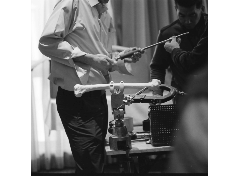
Anatomical models representing the femur as well as its common fractures facilitate residents’ hands-on training in nail assembly, fixation, locking, and other surgical techniques using the femoral nail system.
Surgical Techniques
Below are ways anatomical models help residents train with a retrograde femoral nail system for treating femoral fractures:
Guide Wire Entry and Placement |
Femur models help residents visualize the intercondylar notch as the entry point. Using foam models, residents can practice using the femoral nail system's entry reamer. Proper insertion of guidepin and placement of guidewire using manual force can be practiced repetitively to enhance procedural precision. |
Reduction |
Femur fracture models help demonstrate proper reduction techniques using pull traction. Foam models facilitate practice in placing guidewire through the fracture site using the femoral nail system. Instruction can emphasize techniques in dealing with difficult fractures such as the use of special fracture reducers and pins to enable wider reaming. |
Reaming |
Foam models help familiarize residents with different tool sizes used to ream the intramedullary canal. Best practices such as reaming in increments and avoiding reamer head incarceration can be further discussed. |
Nail Assembly |
Residents can use a femur model to test their proficiency in properly assembling a retrograde nail in preparation for fixation. Manipulation of femoral nail system tools such as the driver nose, targeting arm, and drills can also be practiced on a foam femur model. |
Nail Insertion and Fixation |
Foam models let surgeons acquire proper femur fixation techniques through repetitive practice. These include holding the nail's handle instead of the targeting guide during insertion. Another technique is to carefully advance the nail to avoid the development of new fracture lines in the bone. The use of a mallet or manual force can be tried on foam models during fixation. Proper countersinking of the nail using the femoral nail system guide can also be emphasized. Best practices such as seating the nail fully and using a lateral radiograph to assess insertion depth can be discussed using femur models. |
Screw Placement |
Again, foam models help test residents' proficiency in choosing the right screw length, assembling, and inserting the screw using the femoral nail system. Foam models are suitable for practicing drilling and screw placement. The use of different drill bits and screw sizes can be demonstrated on the model. Techniques such as quick drill removal and screw insertion, tracking of the screw head, and evaluation of the procedure can be demonstrated using models. |
Locking |
Freehand techniques for proximal locking can be practiced on femur models. The use of targeting devices to lock the nail can also be demonstrated. Foam cortical models are suitable for drilling when practicing proximal locking. Proper use of femoral nail system targeting tools and guides during locking can be demonstrated on plastic or foam femur models. |
Nail Removal |
After successful union, femoral nails often need to be removed. Foam models again assist in training residents in proper extraction using a femoral nail system. Best practices such as preventing nail rotation, use of guide wire, and proper extraction technique can be demonstrated on a foam femur model. |
Femur Models
Different femur models serve different purposes in resident program training.
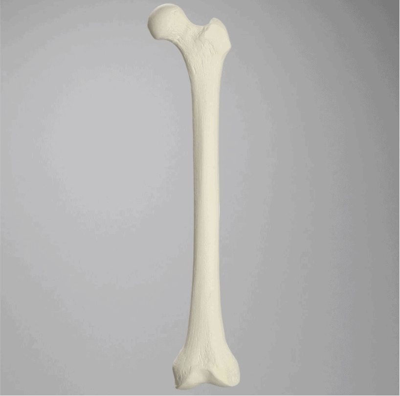
Femur, Foam Cortical, Left, Medium
Foam models are made for easy cutting. This model includes cancellous inner material.
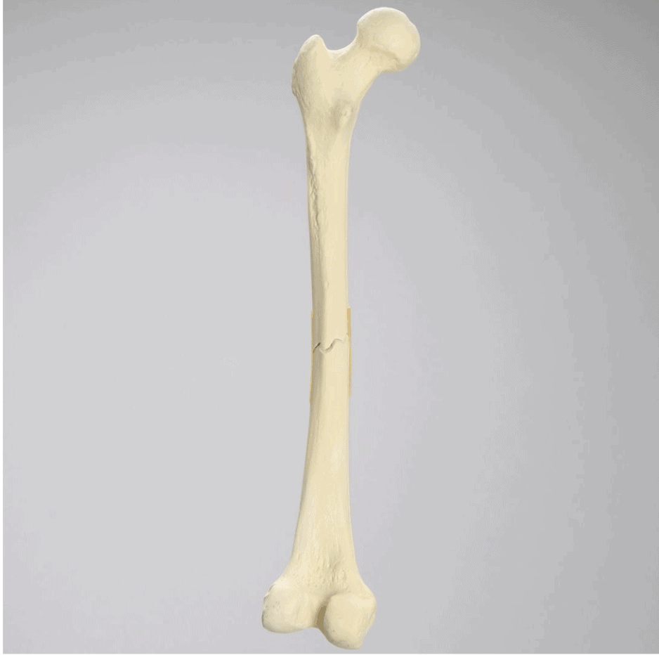
Femur with Transverse Fracture, Foam Cortical, Left, Small
This model includes a midshaft transverse fracture reattached with latex bands.
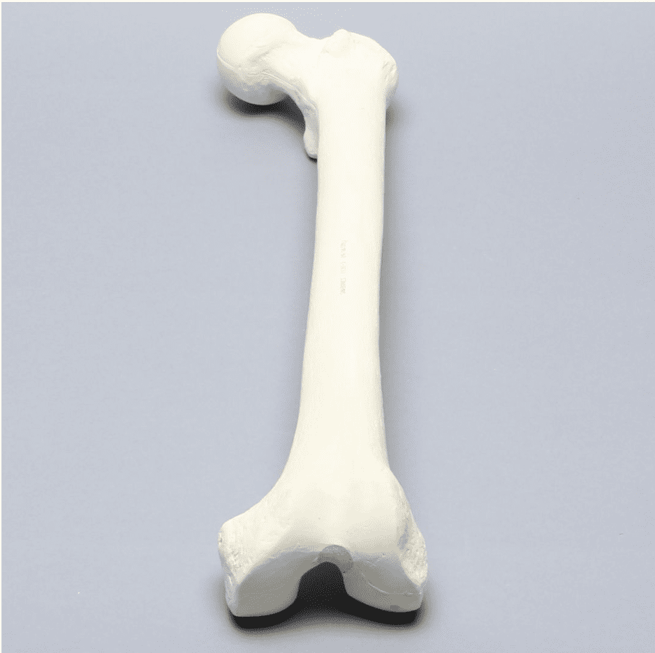
Femur with 17 mm Trochanteric Entry Site For IM Nail
This model is designed for intramedullary nailing applications. It has a canal diameter and greater trochanteric entry side of 17 mm.
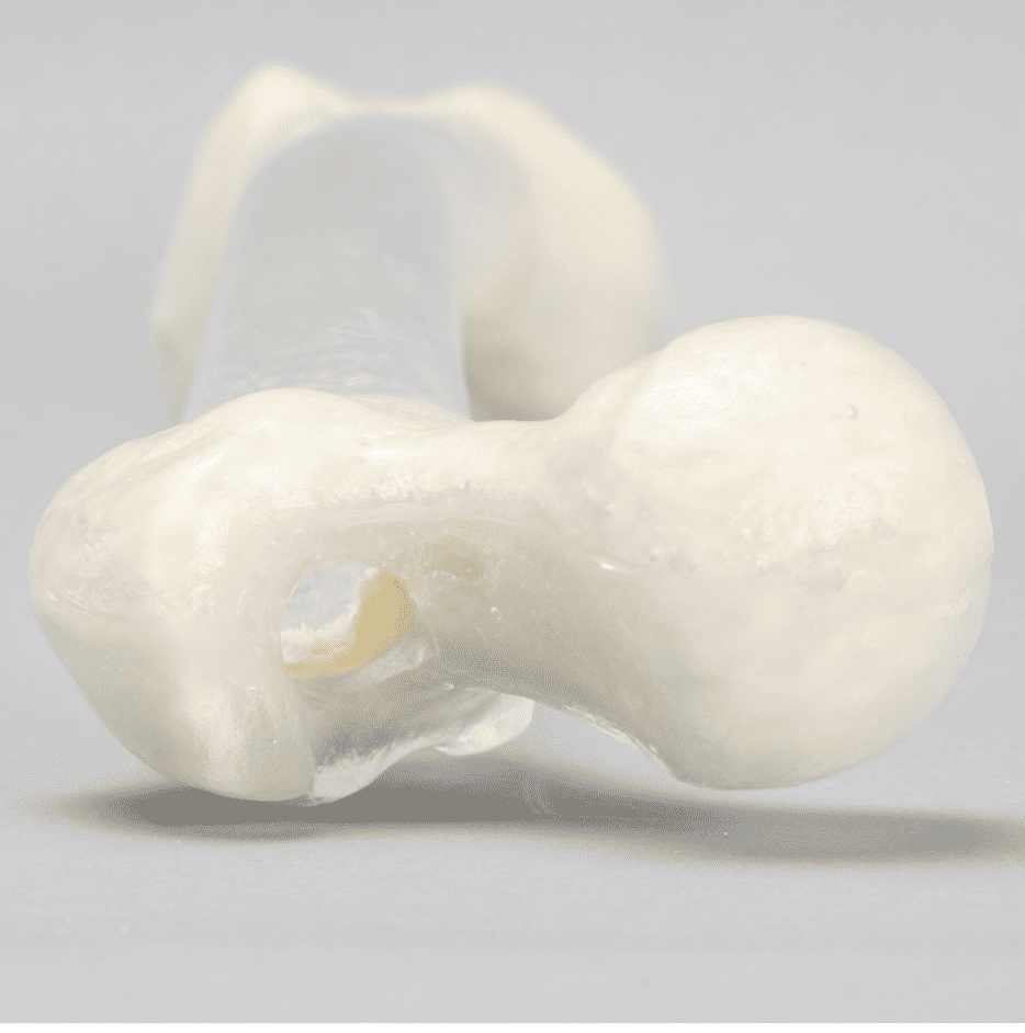
Femur, Transparent with 14 mm Canal, Left
Another model made specifically for intramedullary nailing applications. This one has a transparent canal and proximal entry site of 14 mm.
In summary, anatomical models help residents practice retrograde femoral nail system surgical techniques. These include guide wire entry and placement, fracture reduction, reaming, nail assembly and insertion, screw placement, locking, and nail removal. Foam models are suited for drilling practice while specially designed models are best for intramedullary nailing.
High-Quality Femur Models for Femoral Nail System Training
Sawbones specializes in manufacturing high-quality bone models. Our collection includes foam cortical femur models, plastic cortical femur models, femur models with fractures, models with non-union and malunions, and models for intramedullary nailing among others.
Sawbones offers the best-in-class femur models for training with a femoral nail system. For more information on our offerings or to talk about custom training models, contact us at 206-463-5551.

Work with the industry leader in medical training products!
For four decades, Sawbones has offered the best medical training products for doctors, educators, sales specialists and more. Contact us to learn more about our high-quality models!
1 (206) 463-5551Contact Us!


