Forearm fractures involving the radius and ulna constitute over 40% of pediatric fractures, most of which damage the wrist. In adults, too, distal radius fractures are the most common injury due to the tendency to break one's fall with the hand. Radius and ulna bone model sets help orthopaedic students master the necessary visualization and procedural skills to ensure the success of reduction and fixation treatments.


Typical Radius and Ulna Fractures 

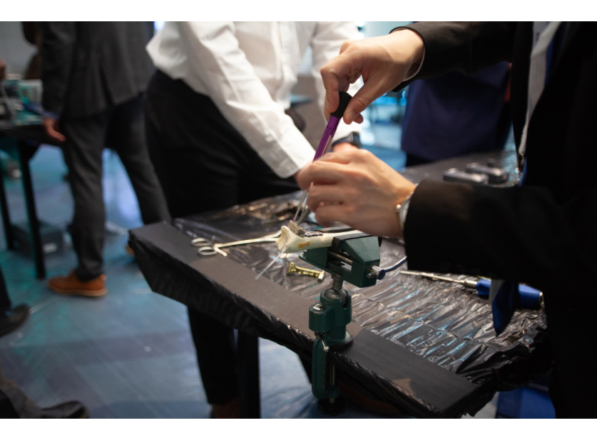
Correct anatomical reduction and stable fixation are essential to restore the function of the arm and hands in the event of significant forearm fractures. Medical schools and training institutions benefit greatly from complementing expert instruction with high quality bone models for hands-on training. Below are ways bone models improve instruction and procedural practice for common radius and ulna fractures.


Fracture Types

Bone Model Training

Distal Radius Fractures

  • Bone models with soft tissue illustrate not only skeletal injury but also possible soft tissue injuries such as ligament and fibrocartilage damage.
  • Bone models help orthopaedic trainees perfect closed reduction percutaneous pinning (CRPP) techniques, which have a very high chance of success in treating distal radius fractures if properly executed.
  • Medical students are trained in the do’s and don’ts of proper immobilization of the wrist ex. Avoid ulnar deviation and excessive flexion.
  • Models provide students with hands-on training with volar and dorsal approaches in open reduction and internal fixation (ORIF) techniques. 
  • Models aid visualization, especially concerning essential nerves and tendons such as the EPL tendon, ulnar nerve, superficial radial nerve, and others. 

Midforearm Fractures


  • Operative techniques for shaft fractures of the radius or ulna are critical to ensure the integrity of anatomic relationships.

  • Poor technique is almost certain to result in delayed union and bowing deformities. Bone models help students rationalize the proper surgical treatment of midforearm fractures. 
  • Models help students practice ORIF methods commonly used to treat shaft fractures of the radius and ulna. Techniques that can be practiced on appropriate bone models (see next section) include compression plating, ORIF with bone grafting, and IM nailing.
  • Full arm models help students practice the application of a functional brace or Muenster cast to control the rotation of the forearm.
  • Special bone models assist students in hands-on training of ulnar nail insertion, proper plate fixation, bridge plating, and bone grafts.
  • Bone models allow students to build dexterity in handling instruments like lobster claw reduction clamps. Reduction techniques such as the application of longitudinal traction and rotation using these instruments can be repetitively practiced. 

Radial Head Fractures

  • Radial head fractures are among the most common elbow fractures resulting from falls on an outstretched hand (FOOSH). Again, models help students explore bone fractures and related injuries to surrounding soft tissue, such as lateral collateral ligament and medial collateral injury. Associated skeletal injuries such as elbow dislocation and carpal fractures can also be demonstrated on bone models with soft tissue.
  • Optimal areas for hardware placement on the radial head can be emphasized with detailed anatomical models.
  • Specifically designed bone models help students practice ORIF techniques such as partial excision of bone fragments, complete radial head excision, athroplasty of displaced comminuted fractures, and special nail reduction. 
  • Surgical approaches to the radial head (Kocher vs Kaplan) can be practiced on appropriate bone models.  Visualization advantages of both approaches and respective risks to nearby nerves and ligaments can be emphasized.
  • Fixation techniques, including implants and plate placement, can be practiced hands-on on unique bone models. 

Growth Plate/Physeal Fractures 

  • Physeal considerations for pediatric patients undergoing surgical treatment can be excellently demonstrated by bone models. For example, pediatric bone is more elastic and exhibits unique fracture patterns such as buckle and greenstick fractures. Open physes also possess greater potential for remodeling bone deformities. Bone models can illustrate such points to establish treatment principles in pediatric surgery.
  • Detailed bone models demonstrate physeal arrest and related complications, including ulnocarpal impaction, DRUJ injury, and TFCC injuries. 
  • Bone models emphasize the importance of proper reduction techniques to avoid growth arrests and deformities in young patients. Instructors may use bone models to demonstrate gentle but firm pressure best used to reduce physeal fractures. Instructors may further emphasize the importance of limiting reduction treatment to one attempt (preferably within a week of the injury) to avoid the risk of growth arrest.


Radius and Ulna Bone Model Choices

Different bone models serve different training purposes. Below are several bone model options designed for visualization and hands-on training of reduction techniques. 

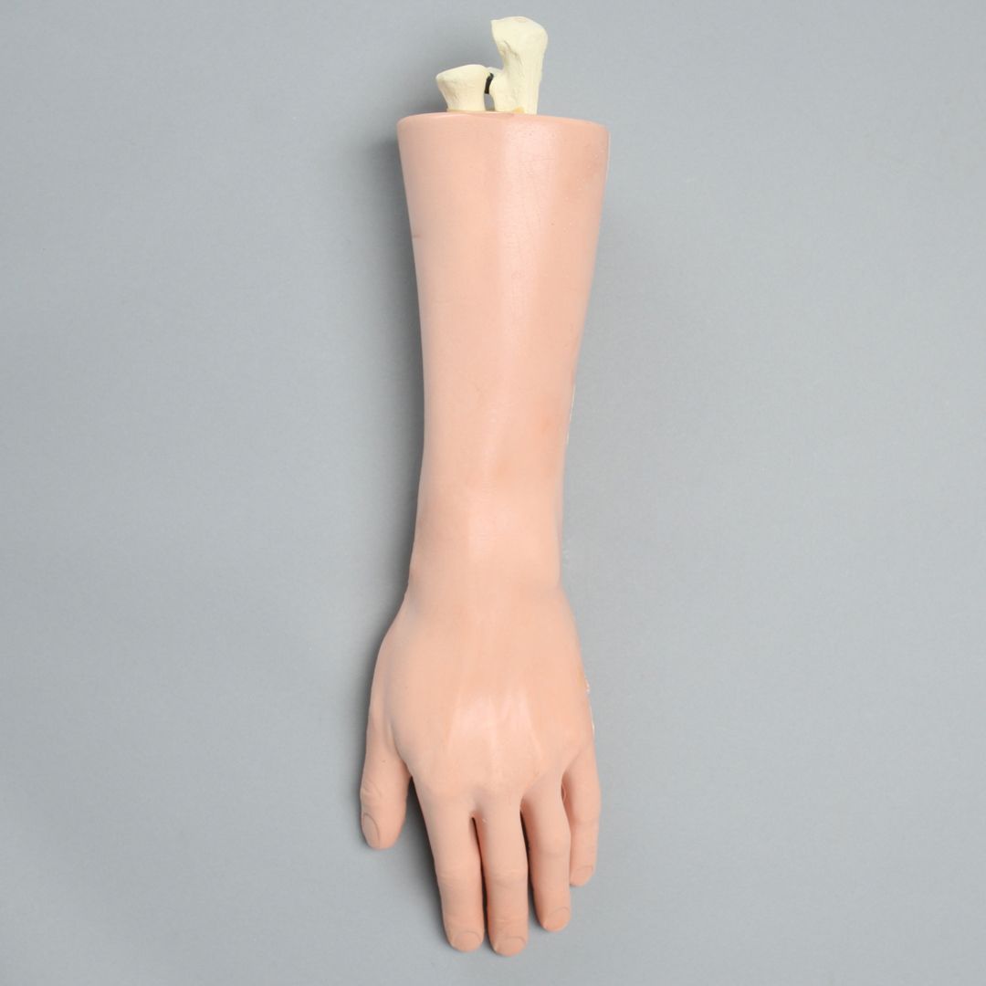
Bone models with soft tissue are useful for hands-on training in open reduction and fixation procedures. 

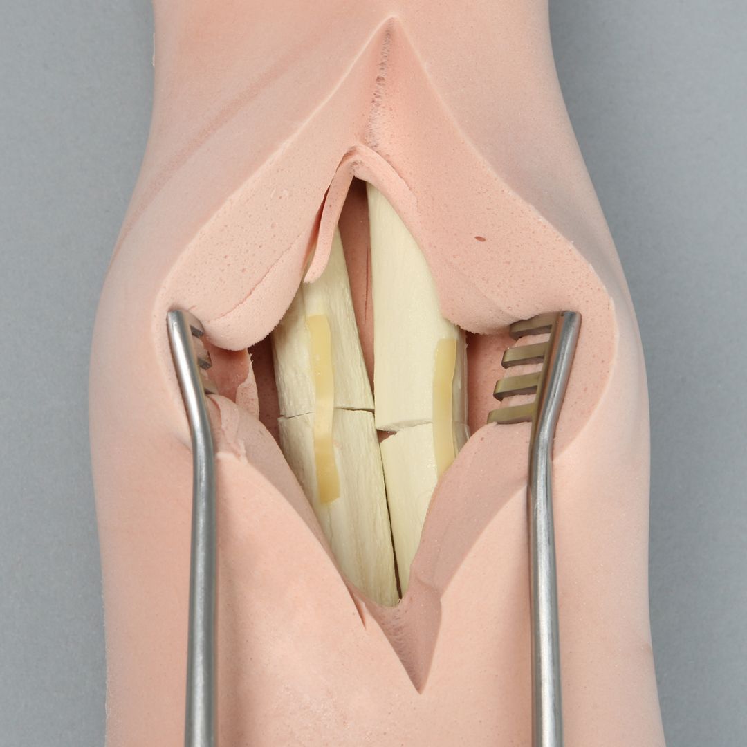
Radius and Ulna Bone Model with Soft Tissue

Hand and Wrist with Midshaft Fractures, Encased

Hand with Midshaft Fractures, Encased  Wrist with Midshaft Fractures, Encased

Foam-based bone models are conducive to drilling, screwing, and plate fixation techniques. 

Hand/wrist Model

Hand and Wrist with Three-Part Fracture, Foam Cortical

Hand and Wrist with Three-Part Fracture, Foam Cortical

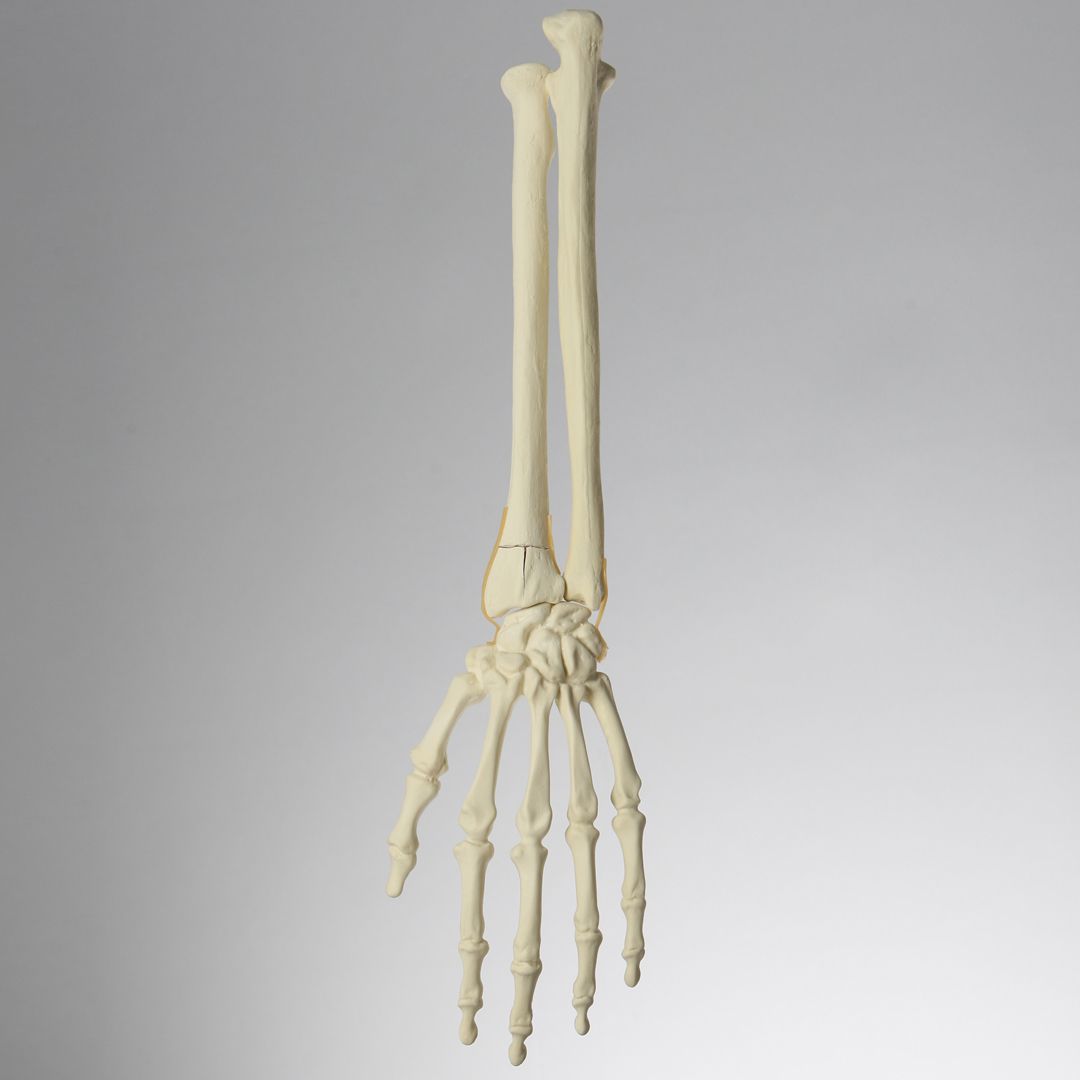
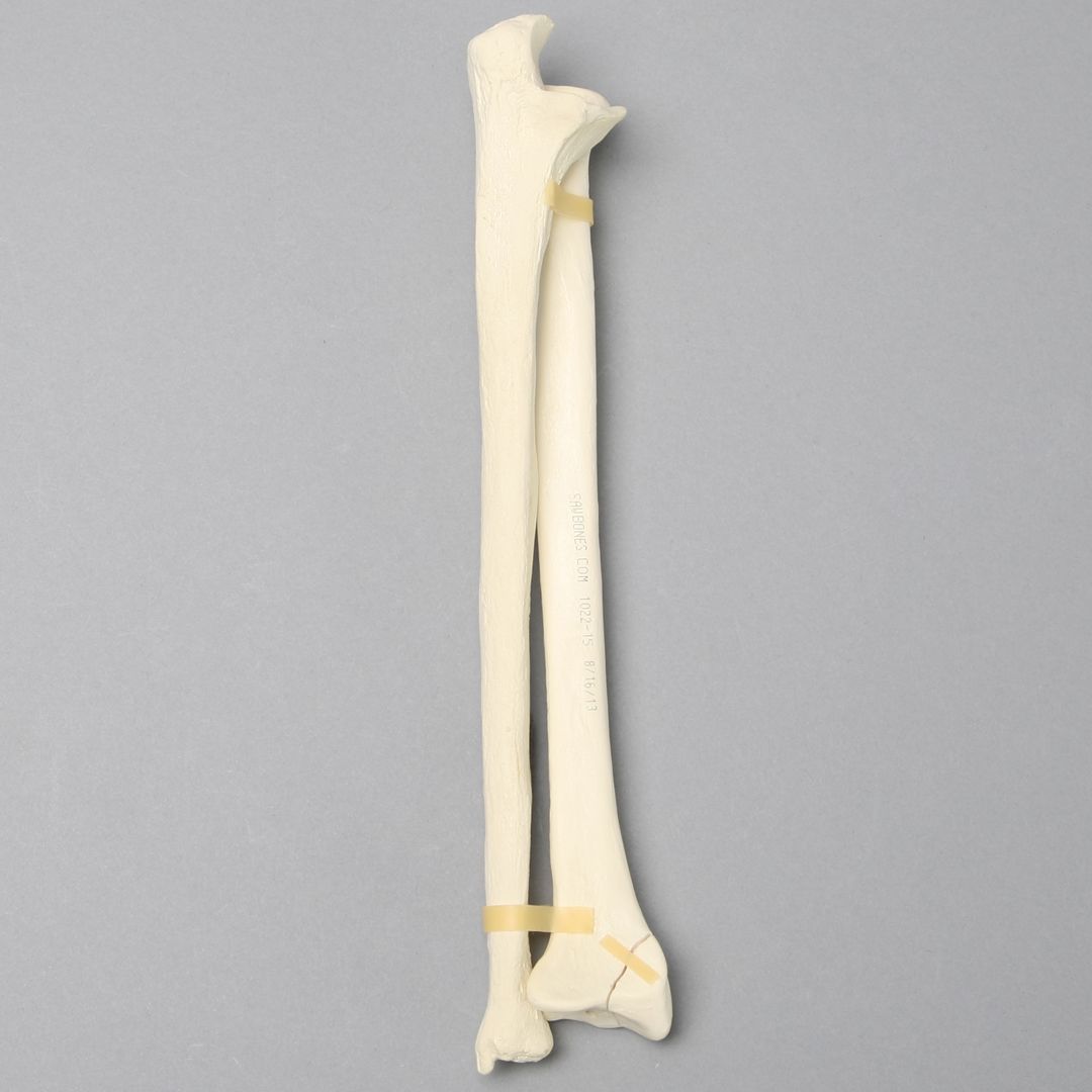
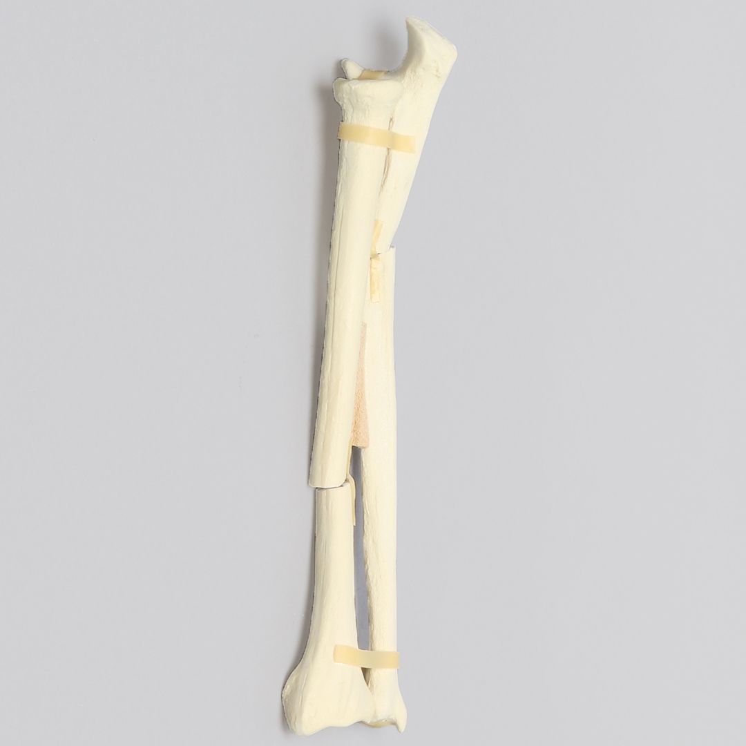
Bone models can also depict single and multiple fractures to further illustrate common injuries. Again, foam models offer excellent hands-on training for treating bone fractures such as plate fixation, nailing, drilling, and other surgical techniques.

 Single Fracture
Ulna and Radius with Styloid Fracture, Foam Cortical

Ulna and Radius with Styloid Fracture, Foam Cortical

Multiple Fractures

Ulna and Radius with Oblique and Transverse Fractures, Foam Cortical

Ulna and Radius with Oblique and Transverse Fractures, Foam Cortical


In conclusion, radius and ulna bone model sets help orthopaedic trainees master ORIF techniques used for treating common forearm fractures. Bone models with soft tissue are best for hands-on training of open and closed reduction techniques. Foam-based bone models lend themselves to drilling, nailing, cutting, and other surgical procedures. Specific sets that depict single and multiple fractures aid remarkably in student familiarization with common radius and ulnar fractures. Finally, full anatomical details offer superior visualization of reduction and fixation treatments. 

Hands-On Bone Models for Orthopaedic Training 

Sawbones offers best-in-class radius and ulna bone model sets for orthopaedic training and medical education. Sawbones is known for originating hands-on workshop models and continues its leadership today in manufacturing anatomical medical training models. We offer different options, including foam, plastic, and radiopaque models. For more information on our offerings or to talk about custom training models, contact us at 206-463-5551.

Technical Sales | Sawbones

If you're seeking something you can't find on our website, our sales team is happy to help. We can either direct you to the right model or provide a free quote on the right custom project to meet your needs. Discover options with our clear bone models, laminated blocks, custom displays, or other machining projects.